. 2018 Apr 16;475(7):1353-1370.
doi: 10.1042/BCJ20180053.

The structure of hydrogenase-2 from Escherichia coli: implications for H2-driven proton pumping

Affiliations

The structure of hydrogenase-2 from Escherichia coli: implications for H2-driven proton pumping

Stephen E Beaton et al. Biochem J. .

Abstract

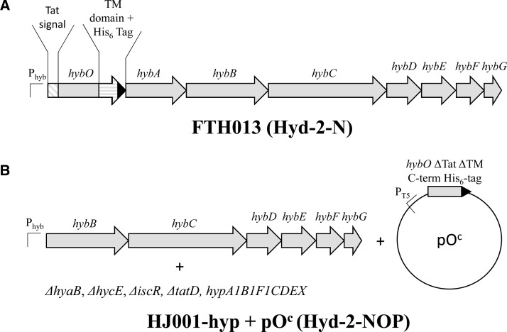
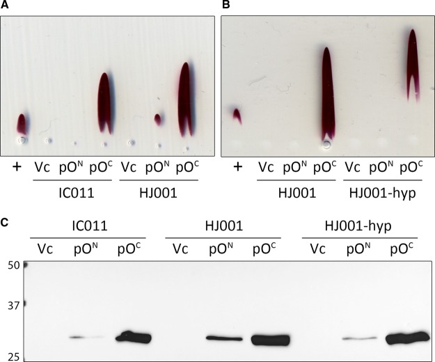
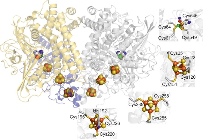
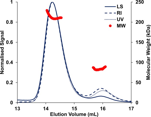
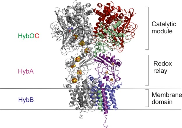
Under anaerobic conditions, Escherichia coli is able to metabolize molecular hydrogen via the action of several [NiFe]-hydrogenase enzymes. Hydrogenase-2, which is typically present in cells at low levels during anaerobic respiration, is a periplasmic-facing membrane-bound complex that functions as a proton pump to convert energy from hydrogen (H2) oxidation into a proton gradient; consequently, its structure is of great interest. Empirically, the complex consists of a tightly bound core catalytic module, comprising large (HybC) and small (HybO) subunits, which is attached to an Fe-S protein (HybA) and an integral membrane protein (HybB). To date, efforts to gain a more detailed picture have been thwarted by low native expression levels of Hydrogenase-2 and the labile interaction between HybOC and HybA/HybB subunits. In the present paper, we describe a new overexpression system that has facilitated the determination of high-resolution crystal structures of HybOC and, hence, a prediction of the quaternary structure of the HybOCAB complex.

Keywords: NiFe hydrogenase; crystallography; electrochemistry; hydrogen metabolism; iron–sulfur clusters; metalloenzymes.


Conflict of interest statement

The Authors declare that there are no competing interests associated with the manuscript.

Figures

Figure 1.
Figure 1.. Schematic representation of the modified hyb operons of strains used in this study.
(A) A representation of the engineered hyb operon of strain FTH013 [5] used for Hyd-2-N production. (B) A representation of the genotype of overexpression host strain HJ001-hyp that carries in-frame ΔhybOA and is transformed with plasmid pOc: this was used to produce Hyd-2-NOP. See Supplementary Table S1 for more information on plasmids and strains.
Figure 2.
Figure 2.. Overproduction of active Hyd-2 using a plasmid-encoded small subunit.
(A and B) Nondenaturing Rocket immunoelectrophoresis was performed using horizontal thin-layers of 1% (w/v) agarose containing 5 µl of anti-Hyd-2 serum buffered with 20 mM barbitone–HCl at pH 8.3. Following electrophoresis, activity staining with 10 mM Tris–HCl (pH 7.5) containing 0.5 mM benzyl viologen and 1 mM tetrazolium red was carried out under a 100% H2 atmosphere. Soluble protein extracts of the FTD674 strain producing truncated Hyd-2 were applied as positive controls (+), and three different host strains IC011 (ΔhyaB, ΔhycE, ΔhybOA), HJ001 (ΔhyaB, ΔhycE, ΔhybOA, ΔiscR), and HJ001-hyp (ΔhyaB, ΔhycE, ΔhybOA, ΔiscR, ΔtatD::hypA1-X) were transformed with either pQE-80L vector (VC) pQE-80L with hybO ΔTat ΔTM N-terminal his-tag (pON), or pQE-80L with hybO ΔTat ΔTM C-terminal his-tag (pOc). (C) Identical soluble extracts used in (A and B) were also analyzed by Western immunoblotting (anti-his-tag antibody) following separation by SDS–PAGE and transfer to the nitrocellulose membrane.
Figure 3.
Figure 3.. The structure of the catalytic core of Hyd-2-NOP containing two copies of the large and small subunits arranged as a dimer of heterodimers ([HybOC]2).
In one heterodimer, the small subunit (HybO) is colored blue and the large subunit (HybC) gold, in the second, both subunits are shown in gray to accentuate the position of the metals centers (displayed as spheres with iron–sulfur clusters colored bronze/yellow and the NiFe center bronze/green). Panels show the coordination of each metal center.
Figure 4.
Figure 4.. SEC-MALLS profile of Hyd-2-NOP in buffer containing detergent (Tris–HCl 20 mM, NaCl 150 mM, DTT 1 mM, and DDM 0.02%, pH 7.2).
The displayed molecular mass corresponds to the protein component of each peak alone (see Methods). UV absorbance was measured at 280 nm. LS: laser scattering; RI: refractive index; MW: molecular mass.
Figure 5.
Figure 5.. Structure of the proximal [4Fe–4S] cluster of HybO with 2Fo–Fc electron density shown as a gray mesh.
(A) In as-isolated crystal, modelling the cluster in a cubane conformation resulted in the appearance of additional FoFc electron density peaks (green mesh) next to the cluster. These peaks were interpreted as an alternate conformation adopted by the cluster upon oxidative damage (B), whereby the Fe atom co-ordinated by Cys25 moves away from the core of the cluster. Rotation of nearby Asp 81 allows the acidic group to co-ordinate the shifted Fe, and an additional water molecule (red sphere) occupies the remaining density next to the damaged cluster. (C) The proximal cluster and associated electron density for reduced Hyd-2 show the cluster is in a single, cubane-like conformation with no uninterpreted FoFc density.
Figure 6.
Figure 6.. The crystal structure of [HybOC]2 is combined with homology models of HybAB to show a proposal for the quaternary configuration of the membrane-bound [HybOCAB]2 complex.
The linker connecting the globular domain of HybO the transmembrane helix has been omitted since it flexibility prohibits modeling its location with any degree of confidence.

References

    1. Sargent F. (2016) The model [NiFe]-hydrogenases of Escherichia coli. Adv. Microb. Physiol. 68, 433–507 10.1016/bs.ampbs.2016.02.008 - DOI - PubMed
    1. Lubitz W., Ogata H., Rüdiger O. and Reijerse E. (2014) Hydrogenases. Chem. Rev. 114, 4081–4148 10.1021/cr4005814 - DOI - PubMed
    1. Fontecilla-Camps J.C., Volbeda A., Cavazza C. and Nicolet Y. (2007) Structure/function relationships of [NiFe]- and [FeFe]-hydrogenases. Chem. Rev. 107, 4273–4303 10.1021/cr050195z - DOI - PubMed
    1. Volbeda A., Martin L., Barbier E., Gutiérrez-Sanz O., De Lacey A.L., Liebgott P.-P. et al. (2015) Crystallographic studies of [NiFe]-hydrogenase mutants: towards consensus structures for the elusive unready oxidized states. J. Biol. Inorg. Chem. 20, 11–22 10.1007/s00775-014-1203-9 - DOI - PubMed
    1. Lukey M.J., Parkin A., Roessler M.M., Murphy B.J., Harmer J., Palmer T. et al. (2010) How Escherichia coli is equipped to oxidize hydrogen under different redox conditions. J. Biol. Chem. 285, 3928–3938 10.1074/jbc.M109.067751 - DOI - PMC - PubMed

Publication types

LinkOut - more resources