. 2018 Mar 19;8(1):4829.
doi: 10.1038/s41598-018-22862-0.

Scoulerine affects microtubule structure, inhibits proliferation, arrests cell cycle and thus culminates in the apoptotic death of cancer cells

Affiliations

Scoulerine affects microtubule structure, inhibits proliferation, arrests cell cycle and thus culminates in the apoptotic death of cancer cells

Klara Habartova et al. Sci Rep. .

Abstract

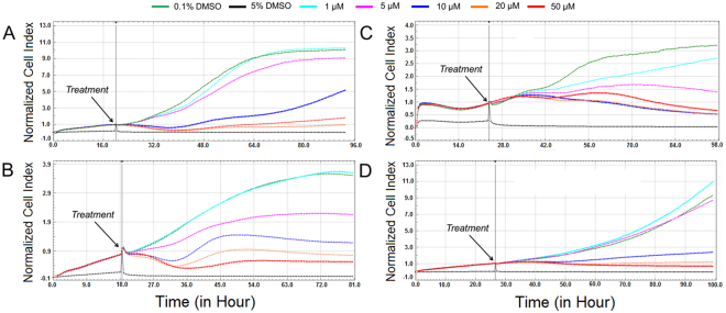
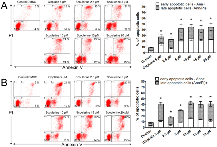
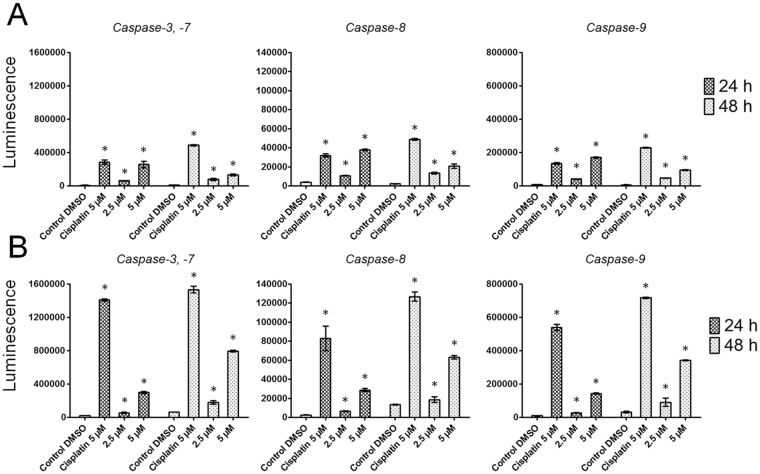
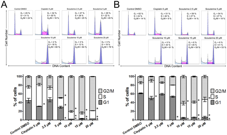
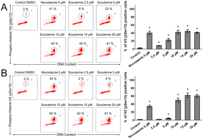
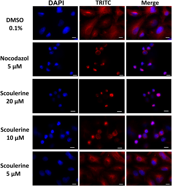
Scoulerine is an isoquinoline alkaloid, which indicated promising suppression of cancer cells growth. However, the mode of action (MOA) remained unclear. Cytotoxic and antiproliferative properties were determined in this study. Scoulerine reduces the mitochondrial dehydrogenases activity of the evaluated leukemic cells with IC50 values ranging from 2.7 to 6.5 µM. The xCELLigence system revealed that scoulerine exerted potent antiproliferative activity in lung, ovarian and breast carcinoma cell lines. Jurkat and MOLT-4 leukemic cells treated with scoulerine were decreased in proliferation and viability. Scoulerine acted to inhibit proliferation through inducing G2 or M-phase cell cycle arrest, which correlates well with the observed breakdown of the microtubule network, increased Chk1 Ser345, Chk2 Thr68 and mitotic H3 Ser10 phosphorylation. Scoulerine was able to activate apoptosis, as determined by p53 upregulation, increase caspase activity, Annexin V and TUNEL labeling. Results highlight the potent antiproliferative and proapoptotic function of scoulerine in cancer cells caused by its ability to interfere with the microtubule elements of the cytoskeleton, checkpoint kinase signaling and p53 proteins. This is the first study of the mechanism of scoulerine at cellular and molecular level. Scoulerine is a potent antimitotic compound and that it merits further investigation as an anticancer drug.


Conflict of interest statement

The authors declare no competing interests.

Figures

Figure 1
Figure 1
Chemical structure and reaction scheme for acylation of scoulerine (1) to 2,9-di-O-acetylscoulerine (2); 2,9-di-O-propionylscoulerine (3) and 2,9-di-O-hexanoylscoulerine (4).
Figure 2
Figure 2
Cytotoxicity of scoulerine (1), (2), (3) and (4) following a single-dose exposure at a concentration of 10 µM. Cell proliferation and viability of Jurkat, MOLT-4, Raji, HL-60, U-937 and HEL 92.1.7 cells measured by using XTT assay 48 h after treatment. Viability is referred to cells treated with 0.1% DMSO (Control DMSO). Data are shown as mean values ± SD of at least three independent experiments. *Significantly different to control (P ≤ 0.05) (A). Dynamic real-time xCELLigence screen of proliferation and cytotoxicity over 62 h. The human A2780 ovarian carcinoma cells in the logarithmic growth phase were treated. The negative control cells were exposed to 0.1% DMSO (vehicle) and 5% DMSO was used as a positive control. The plot is representative of at least three experiments performed (B).
Figure 3
Figure 3
Dynamic real-time monitoring of proliferation and cytotoxicity using the xCELLigence system dedicated to adherent cell lines. Growth kinetics of human A549 lung carcinoma (A), A2780 ovarian carcinoma (B), SK-BR-3 breast adenocarcinoma (C) and MCF-7 breast adenocarcinoma (D) cells treated with scoulerine. Cells treated with 0.1% DMSO were used as vehicle control and 5% DMSO treated cells were used as positive control. The normalized cell index was measured over 72 h. Plots shown are representative of at least three replicate experiments in each case.
Figure 4
Figure 4
The effect of scoulerine on the induction of apoptosis in Jurkat (A) and MOLT-4 (B) leukemic cells. Apoptosis was determined by Annexin V and PI staining 24 h after treatment. Representative histograms of one of three independent measurements are shown. Cells treated with 5 µM cisplatin were used as positive control. The bar graph represents the percentage of early and late apoptotic cells detected by flow cytometry (mean ± SD, n = 3). *Significantly different to control for early and late apoptotic cells (P ≤ 0.05).
Figure 5
Figure 5
The effect of scoulerine on the activity of caspases -3/7, caspase -8 and caspase -9 was determined in Jurkat (A) and MOLT-4 (B) cells. Subsequent to treatment with scoulerine at 2.5 and 5 µM for the indicated times of 24 and 48 h, the cells were analyzed for changes in the activity of caspases-3, -8 and -9 using a luminescent assay. Cells treated with 5 µM cisplatin were used as positive control. *Significantly different to control (P ≤ 0.05).
Figure 6
Figure 6
Analysis of the cell cycle of Jurkat (A) and MOLT-4 (B) leukemic cells 16 h after the application of scoulerine. Cells treated with 5 µM cisplatin were used as a reference compound. The pooled results of 1 of 3 independent experiments are shown. The bar graph represents the percentage of cells in the G1, S, and G2/M phase. Data are presented as mean values ± SD, n = 3. *Significantly different to control (P ≤ 0.05).
Figure 7
Figure 7
The effect of scoulerine treatment on mitotic arrest. The Jurkat (A) and MOLT-4 (B) cells were exposed to scoulerine and 5 μM of nocodazole (an antineoplastic agent that disrupts microtubule function by binding to tubulin used as a reference compound). After 24 h, pSer10 histone H3-positive population (percentages are shown) was quantified by flow cytometry. The figure shows representative flow cytometry histograms depicting Ser10-phosphorylated histone H3 positive cells (gate) in cell cultures. Bars indicate mean ± SD, n = 3. *Significantly different to corresponding control (P ≤ 0.05).
Figure 8
Figure 8
Microscopic images of A549 cells stained with an anti-β-tubulin antibody (red) and counterstained with DAPI (blue). The cells were treated for 24 h with scoulerine or a solvent (0.1% DMSO) as vehicle control. Nocodazole, an antineoplastic agent that disrupts microtubule function by binding to tubulin was used as a reference compound in this assay. Scale bar: 10 µm. Experiments were performed in triplicate using epi-fluorescence microscopy. Photographs from representative chambers are shown. Compared with controls, thicker and denser microtubule bundles were evident in scoulerine-treated cells.
Figure 9
Figure 9
The effect of scoulerine on DNA damage. The alkaline comet assay was used for the detection of single-strand breaks of DNA in Jurkat (A) and MOLT-4 (B) cells at 12 h and 24 h following treatment with scoulerine at 2.5 and 5 µM. The 3% hydrogen peroxide was used as a positive control to verify the comet assay. The dark central bar in each box shows the median. The bottom and top of the box indicate the lower (25%) and upper (75%) quartiles, respectively. Whiskers represent the values within 1.5 interquartile ranges. Values outside this range (outliers) are plotted as solid circles. Extreme values are depicted as asterisks.
Figure 10
Figure 10
Western blot analysis of proteins that regulate cell cycle progression or cell death in Jurkat and MOLT-4 cells after scoulerine treatment for 24 h. Control cells were mock treated with 0.1% DMSO (DMSO). Cells treated with cisplatin at 5 µM were used for positive control in Western blot analysis. These experiments were performed at least three times with similar results and a cropped blot is shown from one representative experiment.

References

    1. Bishayee A, Sethi G. Bioactive natural products in cancer prevention and therapy: Progress and promise. Seminars in Cancer Biology. 2016;40:1–3. doi: 10.1016/j.semcancer.2016.08.006. - DOI - PubMed
    1. Eisenreich WJ, Hofner G, Bracher F. Alkaloids from Croton flavens L. and their affinities to GABA-receptors. Natural Product Research. 2003;17:437–440. doi: 10.1080/1478641031000111516. - DOI - PubMed
    1. Wangchuk P, Keller PA, Pyne SG, Willis AC, Kamchonwongpaisan S. Antimalarial alkaloids from a Bhutanese traditional medicinal plant Corydalis dubia. Journal of Ethnopharmacology. 2012;143:310–313. doi: 10.1016/j.jep.2012.06.037. - DOI - PubMed
    1. Chlebek J, et al. Acetylcholinesterase and butyrylcholinesterase inhibitory compounds from Corydalis cava (Fumariaceae) Natural Product Communications. 2011;6:607–610. - PubMed
    1. Chlebek J, et al. Application of BACE1 immobilized enzyme reactor for the characterization of multifunctional alkaloids from Corydalis cava (Fumariaceae) as Alzheimer’s disease targets. Fitoterapia. 2016;109:241–247. doi: 10.1016/j.fitote.2016.01.008. - DOI - PubMed

Publication types

MeSH terms