. 2018 Mar 19;8(1):4804.
doi: 10.1038/s41598-018-22931-4.

Mechanisms underlying the lack of endogenous processing and CLIP-mediated binding of the invariant chain by HLA-DP84Gly

Affiliations

Mechanisms underlying the lack of endogenous processing and CLIP-mediated binding of the invariant chain by HLA-DP84Gly

Mark Anczurowski et al. Sci Rep. .

Abstract

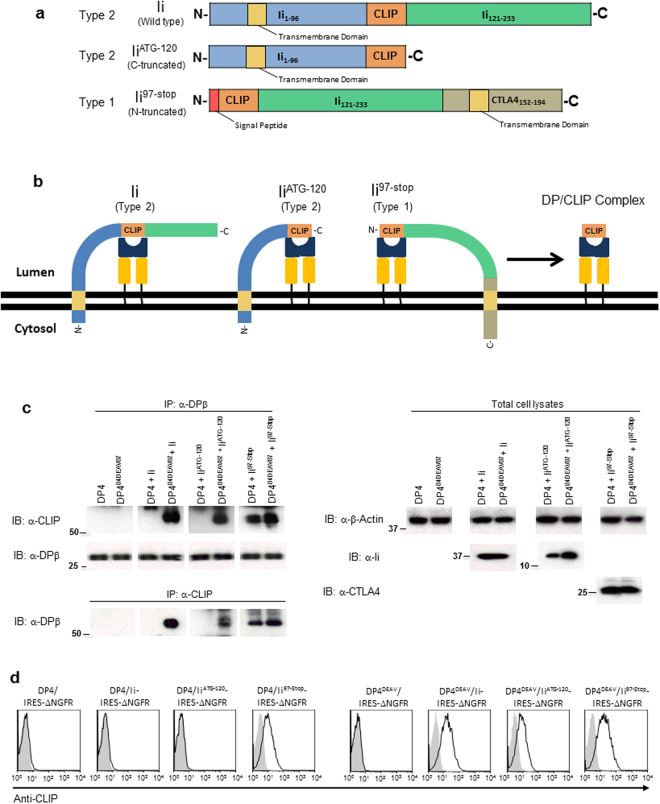
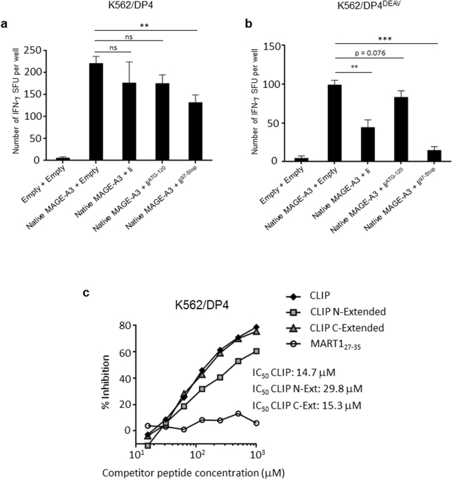
While the principles of classical antigen presentation via MHC class II are well-established, the mechanisms for the many routes of cross-presentation by which endogenous antigens become associated with class II molecules are not fully understood. We have recently demonstrated that the single amino acid polymorphism HLA-DPβ84Gly (DP84Gly) is critical to abrogate class II invariant chain associated peptide (CLIP) region-mediated binding of invariant chain (Ii) to DP, allowing endoplasmic reticulum (ER)-resident endogenous antigens to constitutively associate with DP84Gly such as DP4. In this study, we demonstrate that both the CLIP and N-terminal non-CLIP Ii regions cooperatively generate an Ii conformation that cannot associate with DP84Gly via the CLIP region. We also demonstrate the ability of DP4 to efficiently process and present antigens encoded in place of CLIP in a chimeric Ii, regardless of wild type Ii and HLA-DM expression. These data highlight the complex interplay between DP polymorphisms and the multiple Ii regions that cooperatively regulate this association, ultimately controlling the presentation of endogenous antigens on DP molecules. These results may also offer a mechanistic explanation for recent studies identifying the differential effects between DP84Gly and DP84Asp as clinically relevant in human disease.


Conflict of interest statement

The authors declare no competing interests.

Figures

Figure 1
Figure 1
CLIP is not displayed by HLA-DP molecules encoding the DP84GGPM87 phenotype regardless of HLA-DM expression on K562 cells. Surface class II, CLIP and ΔNGFR expression, along with intracellular Ii and HLA-DM expression, were analyzed on K562 transfected cells by flow cytometry following staining with specific mAbs. K562 cells were retrovirally transduced with DPA1*01:03 (DPA1) and either wild-type or mutated DPB1*04:01 (DPB4), with substitution mutations of the 84–87 region of the DPβ chain made as indicated to generate DP4 and DP484DEAV87 respectively.
Figure 2
Figure 2
The CLIP peptide binds to both DP4 and DP484DEAV87 with greater binding strength than the known DP4-restricted MAGE-A3243–258 peptide. (a) Parental K562 or K562 cells expressing one of DP4 or DP484DEAV87 were pulsed with 100 μM of CLIP, TT947–967, or an equivalent volume of DMSO for 18 hrs. Surface CLIP expression was then analyzed by flow cytometry on the indicated K562 transfected cells following staining with CLIP-specific mAb. (b) A K562 cell-based competitive binding assay was performed using K562-based transfected cells encoding either DP4 or DP484DEAV87, pulsed with graded concentrations of CLIP, MAGE-A3243–258, or MART127–35 peptide in the presence of 2 μM biotin-conjugated reference peptide, HIV ENV31–45. Cells were then stained by phycoerythrin-conjugated streptavidin and analyzed by flow cytometry. Percent inhibition of binding by CLIP peptide was calculated by Mean Fluorescence Intensity (MFI) using the following formula: %inhibition = [1 − (MFI with CLIP peptide/MFI without CLIP peptide)] × 100.
Figure 3
Figure 3
Substitution of the CLIP sequence with the DP4-restricted MAGE-A3243–258 peptide restores multimeric complex formation between Ii and DP84GGPM87. (a) Schema of chimeric Ii constructs encoding MAGE-A3243–258 peptide in place of Ii97–120 (CLIP) sequence. (b,c) HEK293 cells were transiently transfected with the indicated combination of genes and treated with the indicated concentration of the chemical crosslinker DSP for 2 hours. Non-reduced samples were immunoblotted with either anti-Ii (b) or anti-DPβ (c) mAb. Reduced samples were immunoblotted with anti-β-actin mAb. (d,e) HEK 293 cells were transiently transfected with either DP4 or DP484DEAV87 as indicated and either Ii (d) or IiMAGE-A3 (e). Cells were fixed, permeablized and stained using mAbs specific for HLA-DP (red) or Ii (green), then analyzed by confocal microscopy. Inset boxes depict areas shown at a higher magnification. Scale bar in all images represents 10 µm.
Figure 4
Figure 4
DP4 constitutively presents MAGE-A3243–258 peptide derived from chimeric IiMAGE-A3 regardless of the presence of Ii and HLA-DM. (a-h) DP4/MAGE-A3-specific CD4+ T cells were stimulated using the indicated K562-based aAPCs, and IFN-γ responses were measured by ELISPOT analysis. (a) K562 cells stably expressing either DP4 or DP484DEAV87 as indicated were transiently transfected with either IiMAGE-A3 linked with IRES-EGFP, or empty vector (control) encoding IRES-EGFP alone, and used to stimulate T cells. (b) Immunogenicity of stimulator cells was evaluated by stimulating responder T cells with the indicated K562-based aAPCs following pulse with 10 µM of either Tetanus Toxin TT947–967 peptide (control) or MAGE-A3243–258 peptide. (c,d) K562 cells stably expressing DP4, either in the presence or absence of stably expressed Ii and/or HLA-DM as indicated, were transiently transfected with native MAGE-A3 (c), IiMAGE-A3 (d), or empty vector (control) linked with IRES-EGFP, and used to stimulate T cells. (e) Immunogenicity of stimulator cells was evaluated by stimulating responder T cells with the indicated K562-based aAPCs following pulse with 10 µM of either Tetanus Toxin TT947–967 peptide (control) or MAGE-A3243–258 peptide. (f,g) K562 cells stably expressing DP484DEAV87, either in the presence or absence of stably expressed Ii and/or HLA-DM as indicated, were transiently transfected with native MAGE-A3 (f), IiMAGE-A3 (g), or empty vector (control) linked with IRES-EGFP and used to stimulate T cells. (h) Immunogenicity of stimulator cells was evaluated by stimulating responder T cells with the indicated K562-based aAPCs following pulse with 10 µM of either Tetanus Toxin TT947–967 peptide (control) or MAGE-A3243–258 peptide. Transient transfection efficiencies were normalized by EGFP expression as measured by flow cytometry (Supplementary Figure 3). The data shown represent the mean ± SD of experiments performed in triplicate. Results are representative of at least three independent experiments. ns: not significant; *p < 0.05, **p < 0.01, ***p < 0.001 by unpaired, two-tailed Welch’s t-test.
Figure 5
Figure 5
Truncation of N-terminal non-CLIP Ii region(s) restores CLIP-mediated Ii-DP association with DP4. (a) Schema depicting Ii C-terminal (IiATG-120) and N-terminal (Ii97-Stop) truncation mutants compared to wild-type Ii. Note that to generate Ii97-Stop, the transmembrane segment of CTLA4 was fused to the C-terminus of Ii to maintain membrane tethering. (b) Schema depicting Ii truncation mutants compared to wild-type Ii when membrane embedded. Addition of CTLA4 transmembrane region converts Ii97-Stop to a type I transmembrane protein, and consequently the CLIP sequence in each construct is situated on the same side of the ER membrane allowing access to class II as depicted. (c) K562 cells stably expressing the indicated class II allele were transiently transfected with wild-type Ii, IiATG-120, Ii97-Stop or empty vector (control). Lysates were then harvested from these cells, and non-reduced samples were immunoprecipitated and immunoblotted with the indicated mAbs. (d) K562 cells were stably transduced with the indicated combination of genes and then stained using anti-NGFR and anti-CLIP mAbs. The data shown are gated on ΔNGFR+ cells. Full, uncropped blots are provided in Supplementary Fig. 4.
Figure 6
Figure 6
The N-terminal Ii region(s) inhibit CLIP-dependent binding to DP4 in vivo and in vitro. (a,b) DP4/MAGE-A3-specific CD4+ T cells were stimulated using the indicated K562-based aAPCs, and IFN-γ responses were measured by ELISPOT analysis. K562 cells stably expressing DP4 (a) or DP484DEAV87 (b) were transiently transduced with either the empty vector or full-length native MAGE-A3, and one of wild-type Ii, IiATG-120, Ii97-Stop, or the empty vector as indicated and used to stimulate T cells. The transient transfection efficiencies were normalized to EGFP expression as measured by flow cytometry (Supplementary Figure 3). The data shown represent the mean ± SD of experiments performed in triplicate. The results are representative of at least three independent experiments. ns: not significant; **p < 0.01, ***p < 0.001 in unpaired, two-tailed Welch’s t-test. (c) A K562 cell-based competitive binding assay was performed using K562 cells stably expressing DP4. The cells were pulsed with graded concentrations of the CLIP, N-extended CLIP (by five amino acids), C-extended CLIP (by five amino acids), or MART127–35 peptide in the presence of 2 μM of the biotin-conjugated reference peptide HIV ENV31–45. Then, the cells were stained with phycoerythrin-conjugated streptavidin and analyzed by flow cytometry. The percent inhibition of binding by the CLIP peptide was calculated as the mean fluorescence intensity (MFI) using the formula: %inhibition = [1 − (MFI with CLIP peptide/MFI without CLIP peptide)] × 100.

Similar articles

Cited by

References

    1. Wilson NA, et al. Journal of immunology (Baltimore, Md.: 1950) 1998. Invariant chain can bind MHC class II at a site other than the peptide binding groove; pp. 4777–4784. - PubMed
    1. Roche PA, Cresswell P. Invariant chain association with HLA-DR molecules inhibits immunogenic peptide binding. Nature. 1990;345:615–618. doi: 10.1038/345615a0. - DOI - PubMed
    1. Roche PA, Marks MS, Cresswell P. Formation of a nine-subunit complex by HLA class II glycoproteins and the invariant chain. Nature. 1991;354:392–394. doi: 10.1038/354392a0. - DOI - PubMed
    1. Layet C, Germain RN. Invariant chain promotes egress of poorly expressed, haplotype-mismatched class II major histocompatibility complex A alpha A beta dimers from the endoplasmic reticulum/cis-Golgi compartment. Proceedings of the National Academy of Sciences of the United States of America. 1991;88:2346–2350. doi: 10.1073/pnas.88.6.2346. - DOI - PMC - PubMed
    1. Romagnoli P, Germain RN. The CLIP region of invariant chain plays a critical role in regulating major histocompatibility complex class II folding, transport, and peptide occupancy. The Journal of experimental medicine. 1994;180:1107–1113. doi: 10.1084/jem.180.3.1107. - DOI - PMC - PubMed

Publication types

MeSH terms