. 2018 Mar 19;8(1):4827.
doi: 10.1038/s41598-018-23218-4.

Nucleotide-mediated SPDEF modulates TFF3-mediated wound healing and intestinal barrier function during the weaning process

Affiliations

Nucleotide-mediated SPDEF modulates TFF3-mediated wound healing and intestinal barrier function during the weaning process

Sang In Lee et al. Sci Rep. .

Abstract

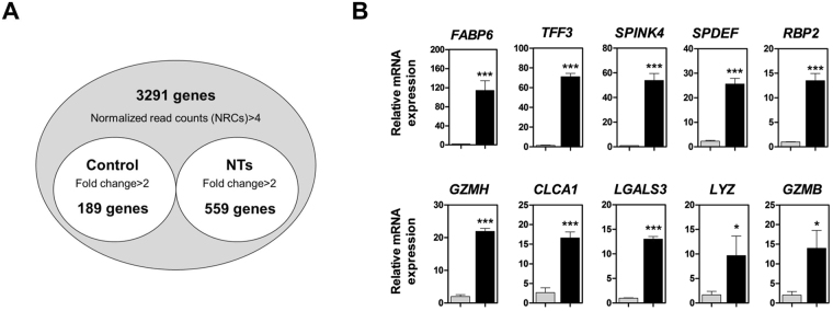
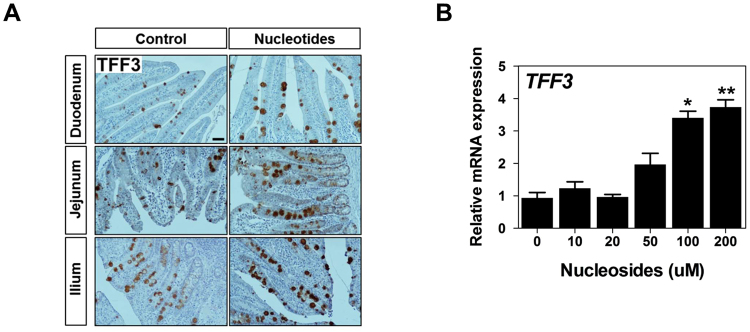
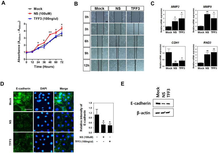
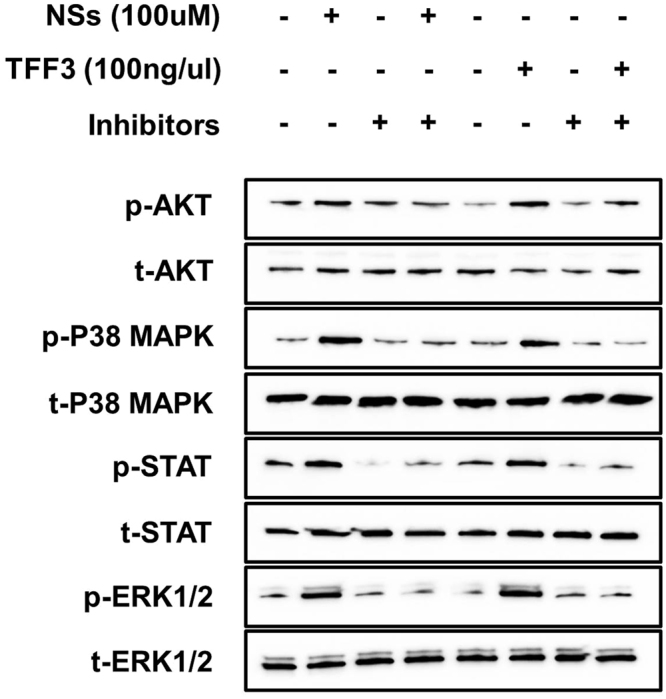
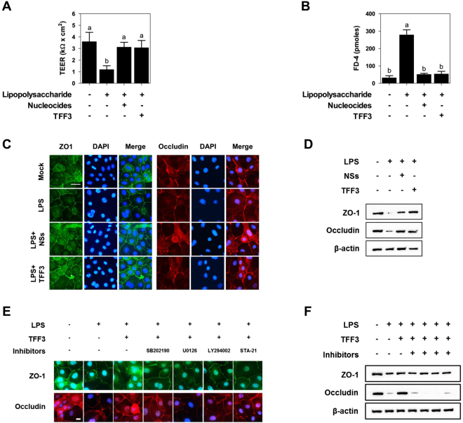
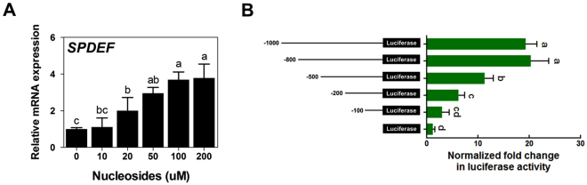
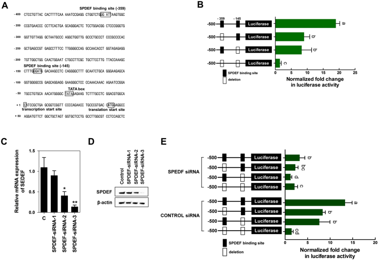
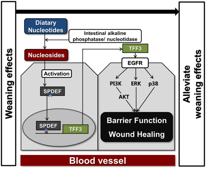
Most alterations during weaning involve physiological changes in intestinal structure and function. Here, we evaluated the molecular mechanisms regulating the effects of nucleotides on weaning. Nucleotide treatment induced Trefoil factor 3 (TFF3) expression and IPEC-J2 cell growth and reduced wound width. Treatment with nucleosides and TFF3 in lipopolysaccharide-challenged IPEC-J2 cells increased intestinal transepithelial electrical resistance and decreased intestinal permeability. Additionally, nucleosides improved intestinal barrier function through induction of TFF3-mediated phosphatidylinositol 3-kinase/Akt, extracellular signal-regulated kinase 1/2, p38, and Janus kinase/signal transducer and activator of transcription signaling pathways. Among selected differentially expressed genes, SAM pointed domain containing ETS transcription factor (SPDEF) expression was elevated by nucleotides in a concentration-dependent manner. Moreover, SPDEF directly regulated TFF3 expression via binding to the promoter. In vivo, nucleotide supplementation improved growth performance, serum stress levels, and intestinal morphology. Our findings provide insights into the molecular mechanisms of intestinal development during weaning in pigs.


Conflict of interest statement

The authors declare no competing interests.

Figures

Figure 1
Figure 1
Gene expression profiling of the small intestine after dietary treatment with NTs for 14 days. (A) Venn diagram illustrations of genes found to be differentially expressed with or without NT treatment in the small intestine. The common genes from quanti-seq experiments were upregulated at least 2-fold (P < 0.05). (B) Quantitative expression analysis of genes that were highly expressed following NT treatment in the small intestine (n = 3). Real-time PCR analysis was conducted in triplicate and normalized to the expression levels of glyceraldehyde 3-phosphate dehydrogenase (GAPDH). Significant differences between control and treatment groups are indicated as ***P < 0.001, **P < 0.01, and *P < 0.05. Error bars indicate standard errors (SEs) of triplicate analyses.
Figure 2
Figure 2
Nucleosides regulated the expression of TFF3. (A) Immunohistochemical staining of TFF3 in the duodenum, jejunum, and ileum with or without NT treatment. Scale bar: 50 μm. (B) IPEC-J2 cells were incubated with varying concentrations of NSs, and ectopically induced expression of TFF3 was analyzed (n = 3). Significant differences between the control (0 μM) and treatment groups are indicated as **P < 0.01. Error bars indicate standard errors (SEs) of triplicate analyses.
Figure 3
Figure 3
Nucleosides regulated TFF3-mediated wound healing. (A) The quantity of viable cells was determined at 12, 24, 36, 48, 60, and 72 h after treatment with NSs (100 μM) and TFF3 (100 ng/µL) using WST-1 assays (n = 3). Significant differences between control and treatment groups are indicated as **P < 0.01 and *P < 0.05. (B) Effects of NSs and TFF3 on migration in IPEC-J2 cells at 0, 3, 6, 9, and 12 h (n = 3). (C) Expression of wound healing-related genes, such as MMP2, MMP9, CDH1, and RND3, was analyzed with or without NSs or TFF3 in IPEC-J2 cells by real-time PCR. (D) Immunocytochemistry of E-cadherin in IPEC-J2 cells was modulated by NSs and TFF3 treatment. Immunofluorescence staining for E-cadherin (green) showed a membranous expression pattern. Nuclei were stained with 4′,6-diamidino-2-phenylindole (DAPI; blue). Fluorescence intensities were quantified using the threshold function of Image J software. (E) Western blotting assay of E-cadherin in IPEC-J2 cells was performed following NS and TFF3 treatment. Significant differences between control and treatment groups are indicated as *P < 0.05. Error bars indicate standard errors (SEs) of triplicate analyses.
Figure 4
Figure 4
Analysis of PI3K/Akt, ERK1/2, p38, and JAK/STAT signaling pathways in IPEC-J2 cells treated with or without NSs or TFF3 (n = 3). IPEC-J2 cells were treated with 100 μM NSs and 100 ng/µL TFF3 for 2 h, and the phosphorylated forms of Akt (Ser473), p38 (Thr180/Tyr182), ERK1/2 (Thr202/Tyr204), and JAK/STAT (Ser727) were detected by western blot analysis. PI3K/Akt (LY2944002), ERK1/2 (U0126), p38 (SB202190), and JAK/STAT (STA-12) were used as inhibitors of corresponding signaling pathways. Whole images of blots are presented in the supplemental information for Fig. 4.
Figure 5
Figure 5
The protective effects of NSs and TFF3 on intestinal barrier function under LPS challenge. (A) Treatment with NSs and TFF3 increased transepithelial-electrical resistance (TEER) under LPS challenge in IPEC-J2 cells (n = 3). (B) Treatment with NSs and TFF3 increased the permeability of fluorescein isothiocyanate-labeled dextrans of 4 kDa (FD-4) under LPS challenge in IPEC-J2 cells (n = 3). Lowercase letters (a,b) indicate significant differences between treatments based on Duncan’s multiple range tests. Error bars indicate standard errors (SEs) of triplicate analyses. Immunofluorescence staining (C) and western blotting (D) showing the effects of NSs and TFF3 on the expression of tight junction proteins in LPS-challenged IPEC-J2 cells. Nuclei were stained with DAPI. Immunofluorescence staining (E) and western blotting (F) indicated that TFF3 improved intestinal barrier function via PI3K/Akt, ERK1/2, p38, and JAK/STAT signaling pathways. LY2944002, U0126, SB202190, and STA-12 were used as inhibitors of the respective signaling pathways. Error bars indicate standard errors (SEs) of triplicate analyses.
Figure 6
Figure 6
Nucleosides regulated the expression of SPDEF. (A) Effects of NSs on the expression of SPDEF in IPC-J2 cells. Significant differences between the control (0 μM) and treatment groups are indicated as *P < 0.05. Error bars indicate standard errors (SEs) of triplicate analyses. (B) Analysis of porcine TFF3 (pTFF3) promoter activity. pTFF3 promoters with different lengths (−1000, −800, −500, −200, and −100) were transfected into IPEC-J2 cells (n = 3). The relative luciferase activity was calculated as the ratio of firefly luciferase to Renilla luciferase. Lowercase letters (a,b,c and d) indicate significant differences between treatments based on Duncan’s multiple range tests. Error bars indicate standard errors (SEs) of triplicate analyses.
Figure 7
Figure 7
Nucleotide-mediated SPDEF induction regulated the expression of TFF3. (A) Nucleotide sequence of the core promoter region for the pTFF3 gene. The numbering of the sequence is relative to the transcription start site. Putative binding sites for the transcriptional factors are boxed and labeled above. (B) Deletion analysis of the pTFF3 promoter. Putative SPDEF binding sites (−359 and −145) were deleted and transfected into IPEC-J2 cells (n = 3). The relative luciferase activity was calculated as the ratio of firefly luciferase to Renilla luciferase. Lowercase letters (a,b and c) indicate significant differences between treatments based on Duncan’s multiple range tests. Error bars indicate standard errors (SEs) of triplicate analyses. qRT-PCR (C) and western blotting (D) indicated that SPDEF expression was suppressed in IPEC-J2 cells using siRNAs. siRNAs were introduced into IPEC-J2 cells by RNAiMAX. Nonspecific siRNA having no complementary sequence in the porcine genome was used as the control. Significant differences between the control and treatment groups are indicated as *P < 0.05 and **P < 0.01. Error bars indicate standard errors (SEs) of triplicate analyses. (E) The effects of SPDEF downregulation on transcription from the pTFF3 promoter. Putative SPDEF binding sites (−359 and −145) were deleted with SPDEF siRNA-3 or control siRNA and transfected into IPEC-J2 cells (n = 3). The relative luciferase activity was calculated as the ratio of firefly luciferase to Renilla luciferase. Lowercase letters (a,b, and c) indicate significant differences between treatments based on Duncan’s multiple range tests. Error bars indicate standard errors (SEs) of triplicate analyses.
Figure 8
Figure 8
Dietary nucleotide supplementation improved growth performance and villus height of the small intestine in weaned pigs. Weaned pigs were randomly allocated into three groups: control (CON), a corn-soybean meal-based control; NT1, CON + 0.5% NTs; and NT2, CON + 1% NTs. (A) Average daily gain (ADG) of pigs in the three groups over time during the experiment. (B) At the end of the feeding trial (day 42), the levels of cortisol, epinephrine, and norepinephrine were determined by ELISA (n = 10). (C) On day 14, the duodenum jejunum, and ileum were sectioned and stained with H&E (n = 24). Lowercase letters (a, b, and c) indicate significant differences between treatments based on Duncan’s multiple range tests. Error bars indicate standard errors (SEs) of analyses.
Figure 9
Figure 9
Schematic illustrating the current working hypothesis regarding the regulation of SPDEF-mediated TFF3 by NT treatment on intestinal development during the weaning process. NTs induced the expression of SPDEF, which regulated TFF3 expression, resulting in modulation of TFF3-mediated wound healing and intestinal barrier function via the PI3K/Akt, ERK1/2, p38, and JAK/STAT signaling pathways.

Similar articles

Cited by

References

    1. Campbell JM, Crenshaw JD, Polo J. The biological stress of early weaned piglets. Journal of animal science and biotechnology. 2013;4:19. doi: 10.1186/2049-1891-4-19. - DOI - PMC - PubMed
    1. Boudry G, Peron V, Le Huerou-Luron I, Lalles JP, Seve B. Weaning induces both transient and long-lasting modifications of absorptive, secretory, and barrier properties of piglet intestine. The Journal of nutrition. 2004;134:2256–2262. doi: 10.1093/jn/134.9.2256. - DOI - PubMed
    1. Le Dividich J, Seve B. Effects of underfeeding during the weaning period on growth, metabolism, and hormonal adjustments in the piglet. Domestic animal endocrinology. 2000;19:63–74. doi: 10.1016/S0739-7240(00)00067-9. - DOI - PubMed
    1. Pacha J. Development of intestinal transport function in mammals. Physiological reviews. 2000;80:1633–1667. doi: 10.1152/physrev.2000.80.4.1633. - DOI - PubMed
    1. Wijtten PJ, van der Meulen J, Verstegen MW. Intestinal barrier function and absorption in pigs after weaning: a review. The British journal of nutrition. 2011;105:967–981. doi: 10.1017/S0007114510005660. - DOI - PubMed

Publication types

MeSH terms

Substances

LinkOut - more resources