. 2018 Nov;32(11):2412-2426.
doi: 10.1038/s41375-018-0082-4. Epub 2018 Feb 28.

High-throughput sequencing of nodal marginal zone lymphomas identifies recurrent BRAF mutations

Affiliations

High-throughput sequencing of nodal marginal zone lymphomas identifies recurrent BRAF mutations

V Pillonel et al. Leukemia. 2018 Nov.

Abstract

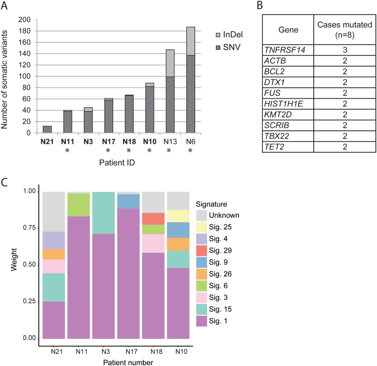
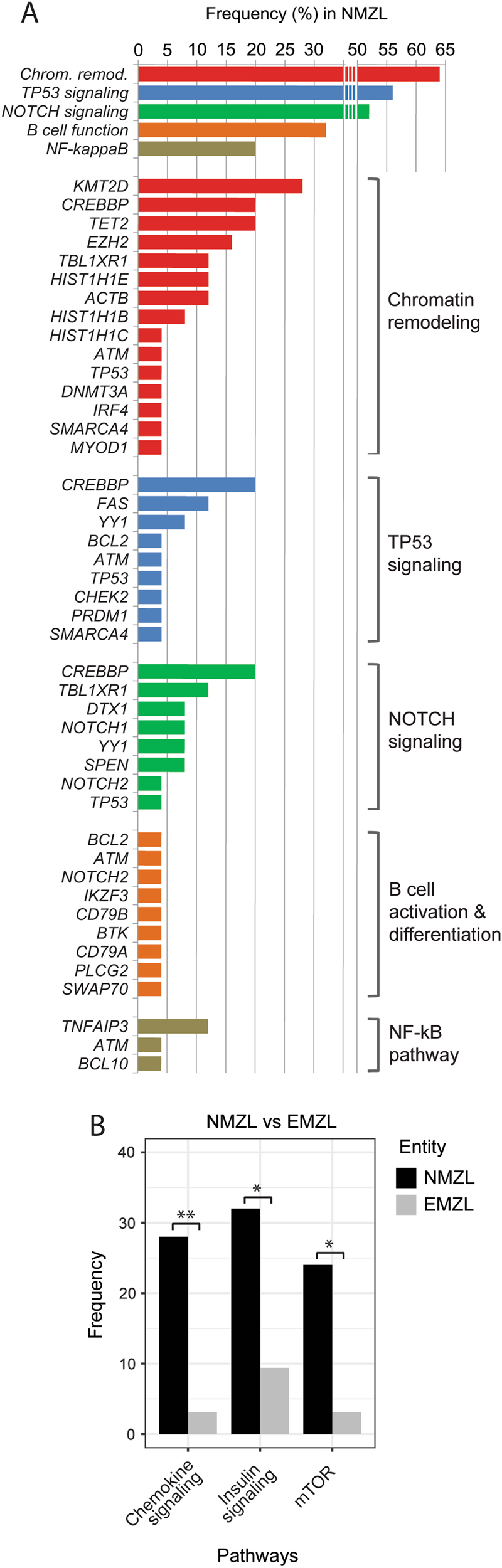
Nodal marginal zone lymphoma (NMZL) is a rare small B-cell lymphoma lacking disease-defining phenotype and precise diagnostic markers. To better understand the mutational landscape of NMZL, particularly in comparison to other nodal small B-cell lymphomas, we performed whole-exome sequencing, targeted high-throughput sequencing, and array-comparative genomic hybridization on a retrospective series. Our study identified for the first time recurrent, diagnostically useful, and potentially therapeutically relevant BRAF mutations in NMZL. Sets of somatic mutations that could help to discriminate NMZL from other closely related small B-cell lymphomas were uncovered and tested on unclassifiable small B-cell lymphoma cases, in which clinical, morphological, and phenotypical features were equivocal. Application of targeted gene panel sequencing gave at many occasions valuable clues for more specific classification.


Conflict of interest statement

The authors declare that they have no conflict of interest.

Figures

Fig. 1
Fig. 1
Whole-exome sequencing of NMZL. a Number and type of single-nucleotide variants (SNVs) and insertions/deletions (InDels) identified in the 8 discovery exomes. Asterisks indicate tumor DNA extracted from fresh frozen tissue. Tumor samples with matched germline tissue are in bold. b Recurrently mutated genes (≥2/8 cases mutated, and selected based on functional annotation and previous data in MZL) detected in the NMZL discovery cohort (n = 8) by whole-exome sequencing. c Mutational signatures based on synonymous and non-synonymous somatic point mutations identified in exomes of NMZL with available paired non-tumoral tissue
Fig. 2
Fig. 2
Landscape of somatic mutations in the studied small B-cell lymphomas. a Number of mutations per sample detected in the different cohorts. Statistical significance by Fisher’s exact test, **p < 0.01; NS not significant. b Heatmap plot showing all non-synonymous mutations detected by targeted high-throughput sequencing in the four small B-cell lymphoma cohorts (NMZL, n = 25; EMZL, n = 32; SMZL, n = 12; and LPL, n = 11). Each row represents a primary tumor grouped according to the assigned subtype. Each column represents a gene ordered left to right in decreasing order of detection frequency. When multiple mutations were present in the same gene, the most damaging mutation is displayed. c Most frequently mutated genes (frequency ≥10%), overall and in each studied entity
Fig. 3
Fig. 3
BRAF is recurrently mutated in NMZL but not in the other studied small B-cell lymphoma entities. a Details of BRAF mutations identified in NMZL. b Representative immunohistochemical staining of a lymph node biopsy of a BRAF mutant NMZL case with the anti-BRAF V600E (clone VE1) antibody. Note positively staining (moderate slightly granular cytoplasmic positivity) tumor cells and adequate internal negative controls. Original magnification: ×240. c Schematic representation of the protein tyrosine kinase domain (Pkinase_Tyr) of the human BRAF protein. Two of the detected non-hotspot mutations (in green) are located in the close proximity to the Val600 hotspot (in red) and are predicted to be damaging in silico
Fig. 4
Fig. 4
Recurrently mutated pathways. a Pathways that are recurrently affected by mutations in NMZL. The bar graphs represent the overall frequency of mutations in each pathway and the frequency of mutations in each gene grouped by pathway. b Differentially mutated pathways between NMZL and EMZL (Fisher’s exact test, *p < 0.05, **p < 0.01)
Fig. 5
Fig. 5
Copy number analysis of NMZL. a Array-comparative genomic hybridization (aCGH) of NMZL cases (n = 22). Red corresponds to gains, blue to losses, and gray to normal (diploid) copy numbers. Trisomies of chromosomes 3, 12, or 18 are highlighted in the boxes. Patients N2, N4, and N25 (indicated by asterisk) had no copy number aberrations (CNAs). b Combined load of somatically acquired genetic lesions identified in the NMZL screening cohort (n = 25), including the numbers of CNA and non-synonymous mutations. Patients samples for which aCGH was not performed are indicated by an asterisk
Fig. 6
Fig. 6
Differentially mutated genes between studied entities. a Genes that are differentially mutated in NMZL compared to EMZL, SMZL, and LPL; heatmap representing alteration frequencies in the differentially mutated genes grouped by predominance in the different subtypes. b Heatmap shows gene mutational frequency comparison results by meta-analysis, which included data from our current cohort and data retrieved from previously published sequencing studies [, , –40]. Only statistically significant enrichments are shown, as determined by Fisher’s exact test, *p ≤ 0.05, **p ≤ 0.01
Fig. 7
Fig. 7
Mutational hints for the classification of unclassifiable small B-cell lymphoma cases. a Heatmap plot showing all non-synonymous mutations detected by targeted high-throughput sequencing in unclassifiable small B-cell lymphomas cases (SBCL, U). The heatmap is clustered by genes and samples with each row representing a primary SBCL, U tumor and each column representing a gene. b Clinico-pathological classification and predicted classification according to the mutation-based evidence of SBCL, U cases

References

    1. Vannata B, Stathis A, Zucca E. Management of the marginal zone lymphomas. Cancer Treat Res. 2015;165:227–49. doi: 10.1007/978-3-319-13150-4_9. - DOI - PubMed
    1. Swerdlow SH, Campo E, Harris NL, Jaffe ES, Pileri SA, Stein H, et al. WHO classification of tumours of haematopoietic and lymphoid tissues. 4th ed. Lyon: International Agency for Research on Cancer; 2017.
    1. Pileri S, Ponzoni M. Pathology of nodal marginal zone lymphomas. Best Pract Res Clin Haematol. 2017;30:50–5. doi: 10.1016/j.beha.2016.11.001. - DOI - PubMed
    1. Traverse-Glehen A, Bertoni F, Thieblemont C, Zucca E, Coiffier B, Berger F, et al. Nodal marginal zone B-cell lymphoma: a diagnostic and therapeutic dilemma. Oncology (Williston Park) 2012;26:92–9. - PubMed
    1. Tadmor T. Nodal marginal zone lymphoma: clinical features, diagnosis, management and treatment. Best Pract Res Clin Haematol. 2017;30:92–8. doi: 10.1016/j.beha.2016.08.026. - DOI - PubMed

Publication types