Review
. 2018 Jun;16(6):341-354.
doi: 10.1038/s41579-018-0003-6.

Autophagy during viral infection - a double-edged sword

Affiliations
Review

Autophagy during viral infection - a double-edged sword

Younho Choi et al. Nat Rev Microbiol. 2018 Jun.

Abstract

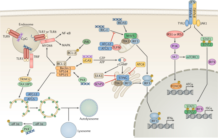
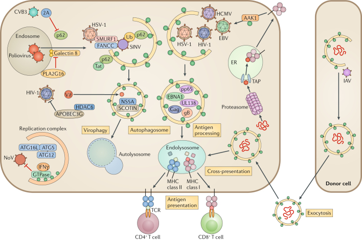
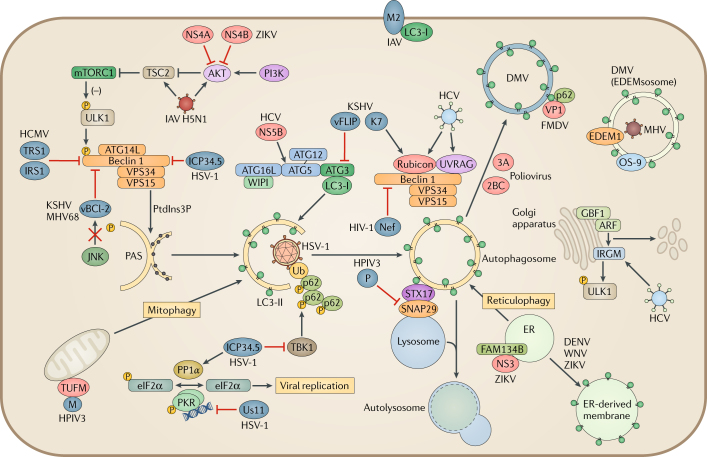
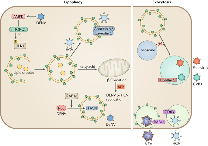
Autophagy is a powerful tool that host cells use to defend against viral infection. Double-membrane vesicles, termed autophagosomes, deliver trapped viral cargo to the lysosome for degradation. Specifically, autophagy initiates an innate immune response by cooperating with pattern recognition receptor signalling to induce interferon production. It also selectively degrades immune components associated with viral particles. Following degradation, autophagy coordinates adaptive immunity by delivering virus-derived antigens for presentation to T lymphocytes. However, in an ongoing evolutionary arms race, viruses have acquired the potent ability to hijack and subvert autophagy for their benefit. In this Review, we focus on the key regulatory steps during viral infection in which autophagy is involved and discuss the specific molecular mechanisms that diverse viruses use to repurpose autophagy for their life cycle and pathogenesis.


Conflict of interest statement

The authors declare no competing interests.

Figures

Fig. 1
Fig. 1. Pattern recognition receptors and autophagy.
In host cells, various pattern-recognition receptors (PRRs) recognize viral pathogen-associated molecular patterns (PAMPs), which leads to the activation of transcription factors and the induction of the interferon (IFN) response. Endosomal Toll-like receptors (TLRs) recognize viral nucleic acids and recruit the adaptor proteins TIR domain-containing adaptor molecule 1 (TRIF) and myeloid differentiation primary response protein MYD88, relaying signals to the nuclear factor-κB (NF-κB) and mitogen-associated protein kinase (MAPK) pathways. Binding to MYD88 or TRIF causes Beclin 1 to dissociate from the B cell lymphoma-2 (BCL-2) inhibitory complex, resulting in the induction of autophagy. TRIF is targeted by selective autophagy by the tripartite motif-containing protein 32 (TRIM32)–TAX1-binding protein 1 (TAX1BP1) complex for degradation. JUN N-terminal kinase (JNK) phosphorylates BCL-2 to initiate Beclin 1-mediated autophagy. During RNA virus infection, retinoic acid-inducible gene I (RIG-I) senses double-stranded (ds)RNA and signals through mitochondrial antiviral-signalling protein (MAVS) to activate interferon regulatory factor 3 (IRF3), leading to the production of IFN. To suppress RIG-I signalling, the autophagy protein 5 (ATG5)–ATG12 complex disrupts the interaction between RIG-I and MAVS. Interferon-induced, dsRNA-activated protein kinase (PKR) phosphorylates eukaryotic translation initiation factor 2 subunit 1 (eIF2α) to induce autophagy. During DNA virus infection, the DNA sensor cyclic GMP–AMP (cGAMP) synthase (cGAS) produces the secondary messenger cGAMP, which activates stimulator of interferon genes protein (STING), IRF3 and IFN expression. Furthermore, cGAS competes with Rubicon for Beclin 1 binding, thus triggering autophagy. Beclin 1 also suppresses cGAMP production and activates ULK1 to phosphorylate STING for its degradation. ATG9 inhibits the aggregation of STING on Golgi apparatus-derived compartments to suppress the DNA sensing. STING undergoes desumoylation and chaperone-mediated autophagy (CMA)-mediated degradation. Finally, extracellular IFN is recognized by the interferon receptor (IFNAR) and activates JAK–STAT (Janus kinase–signal transducer and activator of transcription) signalling. JAK1 and non-receptor tyrosine-protein kinase TYK2 phosphorylate insulin receptor substrate 1 (IRS1) and IRS2, leading to the activation of the PI3K–AKT–mTOR (phosphoinositide 3-kinase–AKT–mechanistic target of rapamycin) pathway and forkhead box 3 transcription factor (FOXO3) as well as the expression of autophagy-related genes. ISGs, interferon-stimulated genes; MDA5, interferon-induced helicase C domain-containing protein 1; SENP2, sentrin-specific protease 2; TBK1, tank-binding kinase 1; TUFM, elongation factor Tu, mitochondrial; VPS34, vacuolar protein sorting 34.
Fig. 2
Fig. 2. Autophagy-mediated antiviral immune responses.
Autophagy targets intruding viruses by directly eliminating them in autophagosomes. Adaptor protein p62 binds to ubiquitin-coated viral particles that are subsequently delivered to the autophagic machinery. For example, p62 binds to Sindbis virus (SINV) capsid protein and mediates selective autophagy by interacting with LC3 through its LC3-interacting region (LIR). Two host factors, E3 ubiquitin-protein ligase SMURF1 and Fanconi anaemia group C protein (FANCC), are involved in the virophagy of SINV and herpes simplex virus type 1 (HSV-1). During hepatitis C virus (HCV) infection, the host endoplasmic reticulum (ER) transmembrane protein SCOTIN associates with HCV non-structural protein 5A (NS5A) and leads to its degradation, restricting HCV replication. Poliovirus infection and membrane rupture are detected by galectin 8, targeting the virus to autophagosomes. Poliovirus uses HRAS-like suppressor 3 (PLA2G16) to escape from autophagic degradation. HIV-1 virion infectivity factor (Vif) is targeted by histone deacetylase 6 (HDAC6) for degradation. The autophagy protein 5 (ATG5)–ATG12–ATG16L1 complex is recruited in the replication complex and restricts norovirus (NoV) replication through interferon-γ (IFNγ)-inducible GTPases. In antigen-presenting cells (APCs), autophagy delivers intracellular and extracellular antigens to the endolysosome, where they are loaded onto major histocompatibility complex (MHC) class II molecules for presentation to CD4+ T cells. EBV nuclear antigen 1 (EBNA1) is the dominant CD4+ T cell antigen and is primarily processed in the autophagosome. HIV-1 Gag is also targeted by the autophagosome for processing. Autophagy regulates the internalization of MHC class I molecules from the cell membrane via AP2-associated kinase 1 (AAK1). UL138 from human cytomegalovirus (HCMV) and gB from HSV-1 are presented on MHC class I molecules in an autophagy-dependent pathway, when antigen peptide transporter (TAP)-dependent presentation is blocked. Autophagy also promotes an alternative pathway of class I presentation called cross-presentation. It modulates trafficking and processing of phagocytosed antigens from the endosome to MHC I, and autophagy induces antigen packaging in donor cells for release to APCs. 2A, protease 2A; CVB3, coxsackievirus B3; EBV, Epstein–Barr virus; IAV, influenza A virus; pp65, 65 kDa phosphoprotein; TCR, T cell receptor; Ub, ubiquitin.
Fig. 3
Fig. 3. Viral manipulation of autophagy.
Viruses interfere with autophagosome formation and fusion with the lysosome. Herpes simplex virus type 1 (HSV-1) neurovirulence factor ICP34.5 directly targets Beclin 1 to block autophagosome formation. ICP34.5 also recruits the phosphatase PP1α to dephosphorylate eukaryotic translation initiation factor 2 subunit 1 (eIF2α) and blocks tank-binding kinase 1 (TBK1)-mediated autophagosome maturation. HSV-1 tegument protein Us11 interacts with interferon-induced, double-stranded RNA-activated protein kinase (PKR) to prevent the phosphorylation of eIF2α and regulates autophagosome formation. Human cytomegalovirus (HCMV) encodes a functional homologue of ICP34.5 called TRS1, which can bind to Beclin 1 and inhibit autophagy. γ-Herpesviruses carry viral homologues of B cell lymphoma-2 (vBCL-2), which attenuate autophagosome formation by interacting with Beclin 1. vBCL-2 evades JUN N-terminal kinase (JNK) phosphorylation, allowing it to bind to Beclin 1 with high affinity. Kaposi’s sarcoma-associated herpesvirus (KSHV) expresses another host protein homologue, vFLIP, that prevents autophagy protein 3 (ATG3) from processing LC3 during phagosome elongation. KSHV K7 interacts with Rubicon and inhibits fusion of the autophagosome with the lysosome. Viruses also control autophagosome formation and remodel vesicles for viral replication. Poliovirus induces autophagy to form a double-membrane vesicle (DMV) for replication. The poliovirus proteins 2BC and 3A increase LC3 lipidation and DMV formation. The foot-and-mouth disease virus (FMDV) capsid protein VP1 uses the adaptor p62 for autophagosome formation. Mouse hepatitis virus (MHV) hijacks the LC3-coated EDEMsosome by accumulating endoplasmic reticulum (ER) degradation-enhancing α-mannosidase-like protein 1 (EDEM1) and OS-9 in DMVs. Flaviviruses (dengue virus (DENV), West Nile virus (WNV) and Zika virus (ZIKV)) use ER-derived membranes for viral replication. ZIKV non-structural protein 4A (NS4A) and NS4B activate autophagy by inhibiting AKT and mechanistic target of rapamycin complex 1 (mTORC1). Furthermore, the viral protease NS3 cleaves reticulophagy regulator 1 (FAM134B) to block reticulophagy so that ZIKV can use the ER as a replication site. Influenza A virus (IAV) M2 binds to and relocalizes LC3 to the plasma membrane. A highly pathogenic IAV H5N1 strain modulates the AKT–tuberin (TSC2)–mTOR pathway to induce autophagy. Human parainfluenza virus type 3 (HPIV3) also directly inhibits the interaction between syntaxin 17 (STX17) and synaptosomal-associated protein 29 (SNAP29) by expressing phosphoprotein P, resulting in the accumulation of autophagosomes. HPIV3 matrix protein M interacts with elongation factor Tu, mitochondrial (TUFM), inducing mitophagy to inhibit the interferon response. Hepatitis C virus (HCV) has multiple targets to regulate the autophagic pathway. The RNA-dependent RNA polymerase NS5B binds to ATG5. By differentially inducing the expression of Rubicon and ultraviolet radiation resistance-associated gene protein (UVRAG), HCV NS4B temporally regulates the autophagic flux to enhance viral replication. Host immunity-related GTPase family M protein (IRGM) is also targeted by HCV for Golgi apparatus fragmentation and viral replication. Viral proteins inducing or inhibiting autophagy are displayed in red and blue, respectively. ARF, ADB ribosylation factor 1; GBF1, Golgi-specific brefeldin A-resistance guanine nucleotide exchange factor 1; IRS1, insulin receptor substrate 1; MHV68, murine γ-herpesvirus 68; PAS, pre-autophagosomal structure; PI3K, phosphoinositide 3-kinase; PtdIns3P, phosphatidylinositol-3-phosphate; Ub, ubiquitin; VPS34, vacuolar protein sorting 34; WIPI, WD-repeat domain phosphoinositide-interacting protein.
Fig. 4
Fig. 4. Viral manipulation of lipophagy and exocytosis.
Dengue virus (DENV) infection activates 5′-AMP-activated protein kinase catalytic subunit α2 (AMPK), which in turn inhibits mechanistic target of rapamycin 1 (mTORC1) to induce lipophagy. Degradation of lipid droplets by DENV-mediated lipophagy contributes to β-oxidation and ATP production in mitochondria, which provides energy for viral replication. DENV viral protease NS3 sequesters fatty acid synthase (FASN) to the sites of DENV replication to increase fatty acid biosynthesis. Hepatitis C virus (HCV) also induces the formation of autophagosome-containing lipid cargos. HCV-induced autophagosomes comprise caveolin 1, caveolin 2 and annexin A2, which promote HCV assembly. Viruses also hijack the exocytosis pathway for the secretion of viral particles. Poliovirus uses phosphatidylserine (PS)-enriched autophagosome-like vesicles for non-lytic release. Coxsackievirus B3 (CVB3) exits cells through autophagosomes that contain the exosome marker flotillin 1. HCV is also released through exocytic pathways that are regulated by autophagy. Enveloped double-stranded DNA viruses replicating in the nucleus obtain their second envelope from endoplasmic reticulum (ER) and Golgi apparatus membranes. Varicella zoster virus (VZV) exits cells with autophagic membranes that contain lipidated LC3 and the endocytosis marker RAB11.

References

    1. Levine B, Mizushima N, Virgin HW. Autophagy in immunity and inflammation. Nature. 2011;469:323–335. doi: 10.1038/nature09782. - DOI - PMC - PubMed
    1. Levine B, Kroemer G. Autophagy in the pathogenesis of disease. Cell. 2008;132:27–42. doi: 10.1016/j.cell.2007.12.018. - DOI - PMC - PubMed
    1. Mizushima N, Yoshimori T, Ohsumi Y. The role of Atg proteins in autophagosome formation. Annu. Rev. Cell. Dev. Biol. 2011;27:107–132. doi: 10.1146/annurev-cellbio-092910-154005. - DOI - PubMed
    1. Mijaljica D, Prescott M, Devenish RJ. Microautophagy in mammalian cells: revisiting a 40-year-old conundrum. Autophagy. 2011;7:673–682. doi: 10.4161/auto.7.7.14733. - DOI - PubMed
    1. Sahu R, et al. Microautophagy of cytosolic proteins by late endosomes. Dev. Cell. 2011;20:131–139. doi: 10.1016/j.devcel.2010.12.003. - DOI - PMC - PubMed

Publication types