. 2018 Mar 19;8(1):4832.
doi: 10.1038/s41598-018-22902-9.

Self-assembling asymmetric peptide-dendrimer micelles - a platform for effective and versatile in vitro nucleic acid delivery

Affiliations

Self-assembling asymmetric peptide-dendrimer micelles - a platform for effective and versatile in vitro nucleic acid delivery

Ganesh R Kokil et al. Sci Rep. .

Abstract

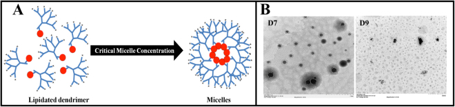
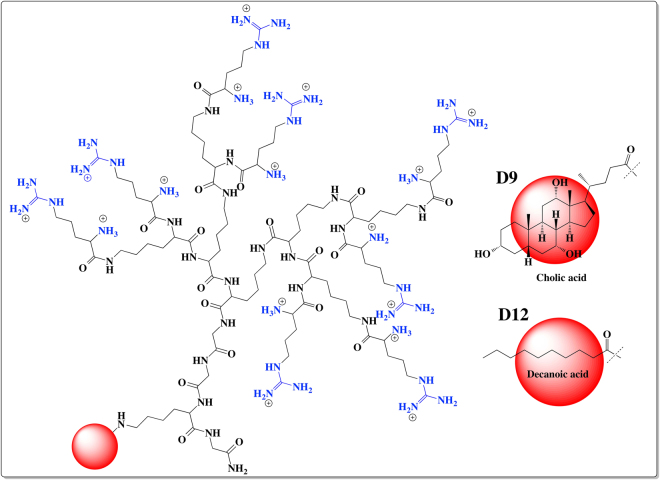
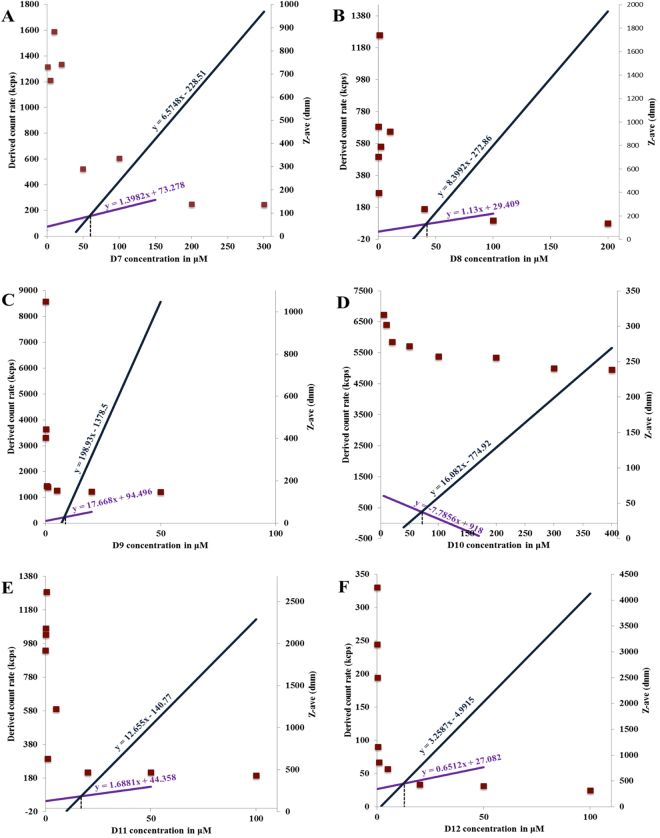
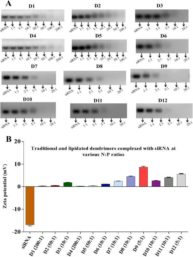
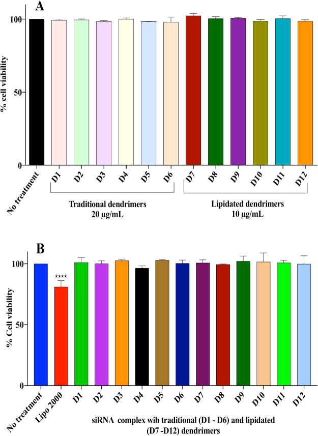
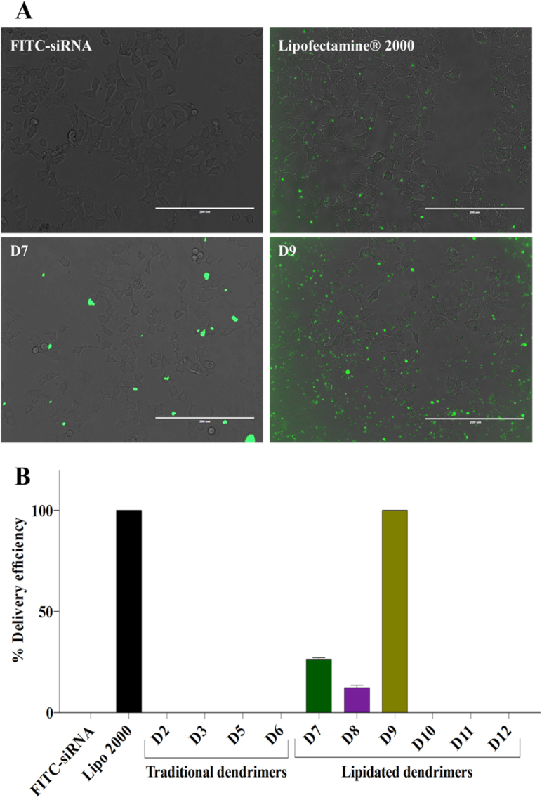
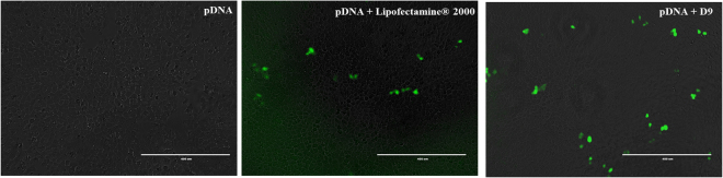
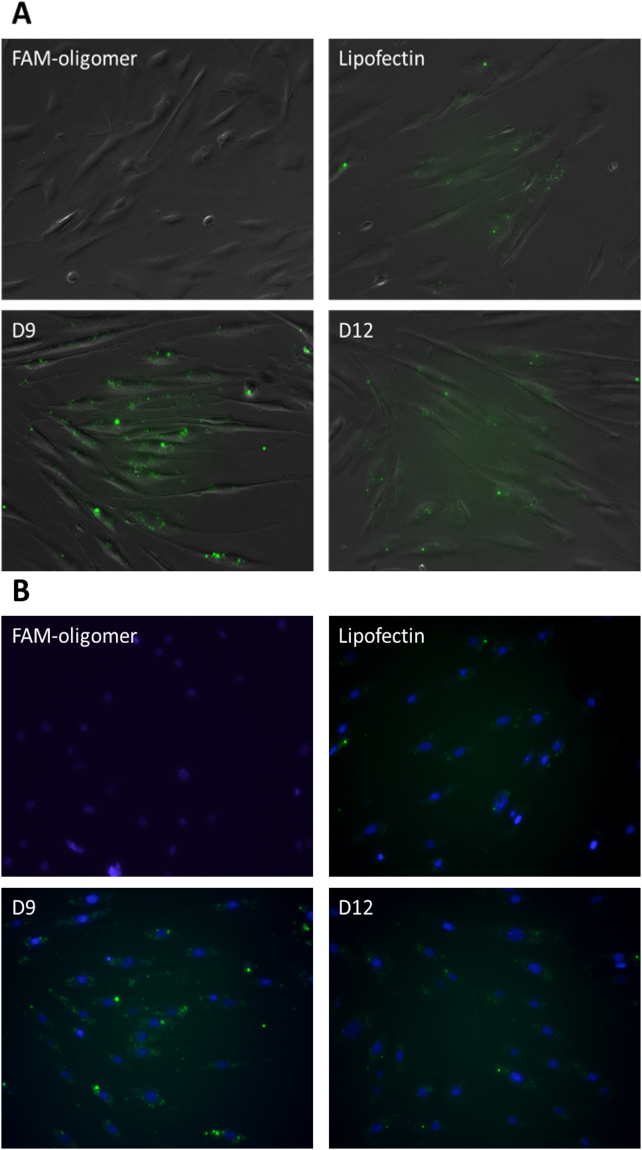
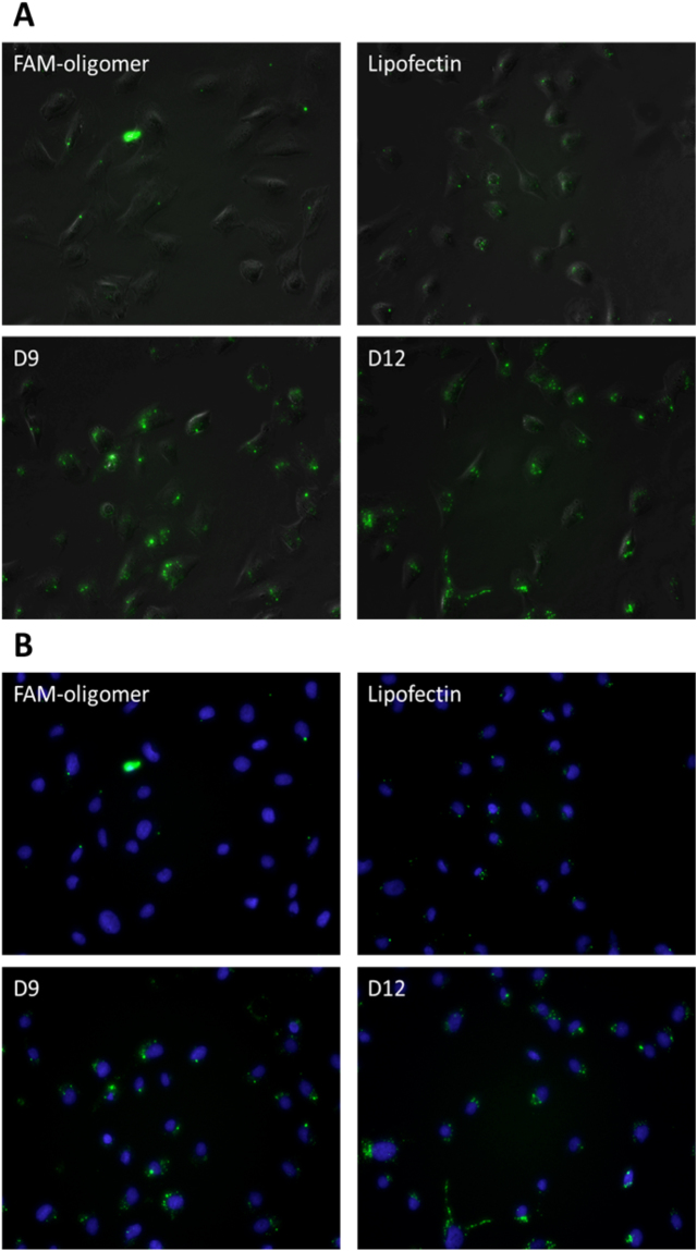
Despite advancements in the development of high generation cationic-dendrimer systems for delivery of nucleic acid-based therapeutics, commercially available chemical agents suffer from major drawbacks such as cytotoxicity while being laborious and costly to synthesize. To overcome the aforementioned limitations, low-generation cationic peptide asymmetric dendrimers with side arm lipid (cholic and decanoic acid) conjugation were designed, synthesized and systematically screened for their ability to self-assemble into micelles using dynamic light scattering. Cytotoxicity profiling revealed that our entire asymmetric peptide dendrimer library when trialled alone, or as asymmetric dendrimer micelle-nucleic acid complexes, were non-cytotoxic across a broad concentration range. Further, the delivery efficiency of asymmetric peptide dendrimers in H-4-II-E (rat hepatoma), H2K (mdx mouse myoblast), and DAOY (human medulloblastoma) cells demonstrated that cholic acid-conjugated asymmetric dendrimers possess far superior delivery efficiency when compared to the commercial standards, Lipofectamine 2000 or Lipofectin®.


Conflict of interest statement

The authors declare no competing interests.

Figures

Figure 1
Figure 1
(A) Schematic representation of a lipidated (red ball) asymmetric peptide dendrimer (blue branches) micelle forming above its critical micelle concentration (CMC) (B) Representative TEM images of D7 and D9, showing morphology characterization of the self-assembled micelles.
Figure 2
Figure 2
Chemical structures of asymmetric peptide dendrimers. 16+ Arg-CA (D9), 16+ Arg-DA (D12).
Figure 3
Figure 3
A comparative plot of intensity of scattered light (left axis) and Z-ave (right axis) as a function lipidated asymmetric peptide dendrimer (D7-D12) concentrations.
Figure 4
Figure 4
(A) N to P ratio for D1 to D6 (traditional) and D7 to D12 (lipidated) asymmetric peptide dendrimers with amine head group. (B) Net surface charge on siRNA and dendriplexes (at optimal N to P ratios) determined using zeta potential (mV) measurements.
Figure 5
Figure 5
siRNA protection by D2 and D5 asymmetric peptide dendrimers.
Figure 6
Figure 6
(A) Evaluation of cell viability on H-4-II-E cells after treatment with traditional and lipidated asymmetric peptide dendrimers. H-4-II-E cells were seeded at 1 × 10⁴ cells per well in 24-well plates and incubated at 37 °C in cell culture medium with 10% FBS for 24 h. Cells were exposed to FITC-siRNA (2 μg/mL), Lipofectamine 2000/FITC-siRNA (2 μg/mL) complex, or asymmetric peptide dendrimers/FITC-siRNA (2 μg/mL) complex in serum-free medium for 4 h. The complex was removed and cells incubated in 10% FBS medium for 24 h and imaged using fluorescence microscopy. Asymmetric peptide dendrimer internalization were visualized by representative bright field and fluorescence microscopy images of cells 24 h post treatment and compared to FITC-siRNA alone, Lipofectamine 2000/FITC-siRNA or Asymmetric peptide dendrimers/FITC-siRNA as indicated. Scale bar represents 200 μm. (B) Assessment of cell viability after exposure to traditional and lipidated asymmetric peptide dendrimers/siRNA complex H-4-II-E cells were exposed for 4 h to serum-free medium (no treatment), traditional (D1 to D6) and lipidated (D7 to D12) asymmetric peptide dendrimers /siRNA complex at various optimized N to P ratios, or to Lipofectamine 2000 /siRNA (0.1 pmol) complex. Cell viability was assessed after 4 h post treatment using the MTS assay. Results are expressed as percentage cell viability compared to untreated control cells and shown as mean ± S.E.M. (n=3 separate experiments), ****p<0.0001 using two way ANOVA.
Figure 7
Figure 7
(A) Delivery efficiency of cationic asymmetric peptide dendrimers determined using FITC-labelled siRNA. H-4-II-E cells were seeded at 1 × 104 cells per well in 24-well plates and incubated at 37 °C in cell culture medium with 10% FBS for 24 h. Cells were exposed to FITC-siRNA (2 μg/mL), Lipofectamine 2000/FITC-siRNA (2 μg/mL) complex, or asymmetric peptide dendrimers/FITC-siRNA (2 μg/mL) complex in serum-free medium for 4 h. The complex was removed and cells incubated in 10% FBS medium for 24 h and imaged using fluorescence microscopy. Asymmetric peptide dendrimer internalization were visualized by representative bright field and fluorescence microscopy images of cells 24 h post treatment and compared to FITC-siRNA alone, Lipofectamine 2000/FITC-siRNA or Asymmetric peptide dendrimers/FITC-siRNA as indicated. Scale bar represents 200 μm. (B) Asymmetric peptide dendrimers delivery efficiency using FITC-siRNA. The asymmetric peptide dendrimer internalization was reported as % FITC-siRNA delivery efficiency, compared to FITC-siRNA alone and Lipofectamine 2000/ FITC-siRNA, quantitated using Image J software.
Figure 8
Figure 8
Delivery efficiency of cationic asymmetric peptide dendrimer D9 determined using pEGFP expression. H-4-II-E cells were seeded at 1 × 10⁴ cells per well in 24-well plates and incubated at 37 °C in cell culture medium with 10% FBS for 24 h. Cells were exposed to pEGFP (1 μg/mL), Lipofectamine 2000/pEGFP (1 μg/mL) complex, or asymmetric peptide dendrimers/pEGFP (1 μg/mL) complex in serum-free medium for 4 h. The complex was removed and cells incubated in 10% FBS medium for 48 h and imaged using fluorescence microscopy. pEGFP expressions were visualized by representative bright field and fluorescence microscopy images of cells 48 h post treatment and compared to pEGFP, Lipofectamine 2000/pEGFP or asymmetric peptide dendrimers/pEGFP as indicated. Scale bar represents 400 μm.
Figure 9
Figure 9
Delivery efficiency of asymmetric peptide dendrimers determined using FAM-ssDNA in H2K mdx cell line. DAOY cells were plated in a 24-well plate 24 hours prior to transfection at 2.5 × 10⁴ cells/well. Cells were transfected with FAM-ssDNA (3.5 μg/mL), Lipofectin®/ FAM-ssDNA (3.5 μg/mL) complex, or asymmetric peptide dendrimer/FAM-ssDNA (3.5 μg/mL) complex in serum-free medium for 24 h. Twenty-four hours after transfection, the cell nucleus were stained with Hoechst for 15 minutes, washed with PBS before asymmetric peptide dendrimers internalization were visualized by representative in (A) bright field and fluorescence microscopy; and (B) UV and fluorescein-based microscopy with nucleus staining.
Figure 10
Figure 10
Delivery efficiency of asymmetric peptide dendrimers determined using FAM-ssDNA in DAOY cell line. DAOY cells were plated in a 24-well plate 24 hours prior to transfection at 2.5 × 10⁴ cells/well. Cells were transfected with FAM-ssDNA (3.5 μg/mL), Lipofectin®/ FAM-ssDNA (3.5 μg/mL) complex, or asymmetric peptide dendrimer/FAM-ssDNA (3.5 μg/mL) complex in serum-free medium for 24 h. Twenty-four hours after transfection, the cell nucleus were stained with Hoechst for 15 minutes, washed with PBS before asymmetric peptide dendrimers internalization were visualized by representative in (A) bright field and fluorescence microscopy; and (B) UV and fluorescein-based microscopy with nucleus staining.

Similar articles

Cited by

References

    1. Tupally, K. R., Kokil, G. R., Thakur, S. S., Singh, P. & Parekh, H. S. in Controlled Release Systems: Advances in Nanobottles and active Nanoparticles (eds J Forcada, A van Herk, & G Pastorin) (Pan Stanford 2015).
    1. Xu H, Li Z, Si J. Nanocarriers in gene therapy: a review. J Biomed Nanotechnol. 2014;10:3483–3507. doi: 10.1166/jbn.2014.2044. - DOI - PubMed
    1. Jain K, Kesharwani P, Gupta U, Jain NK. Dendrimer toxicity: Let’s meet the challenge. Int J Pharm. 2010;394:122–142. doi: 10.1016/j.ijpharm.2010.04.027. - DOI - PubMed
    1. Madaan K, Kumar S, Poonia N, Lather V, Pandita D. Dendrimers in drug delivery and targeting: Drug-dendrimer interactions and toxicity issues. J Pharm Bioallied Sci. 2014;6:139–150. doi: 10.4103/0975-7406.130965. - DOI - PMC - PubMed
    1. Janaszewska A, et al. Cytotoxicity of PAMAM, PPI and maltose modified PPI dendrimers in Chinese hamster ovary (CHO) and human ovarian carcinoma (SKOV3) cells. New Journal of Chemistry. 2012;36:428–437. doi: 10.1039/C1NJ20489K. - DOI

Publication types