. 2018 Mar 19;9(1):1132.
doi: 10.1038/s41467-018-03426-2.

Wnt ligands influence tumour initiation by controlling the number of intestinal stem cells

Affiliations

Wnt ligands influence tumour initiation by controlling the number of intestinal stem cells

D J Huels et al. Nat Commun. .

Abstract

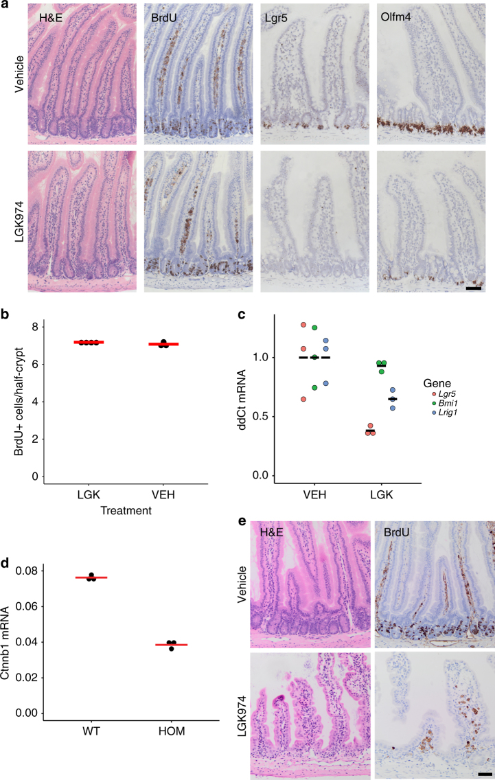
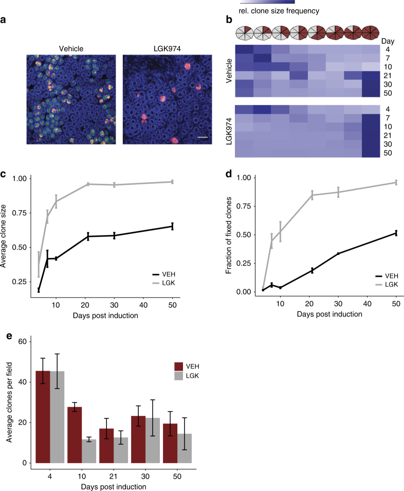
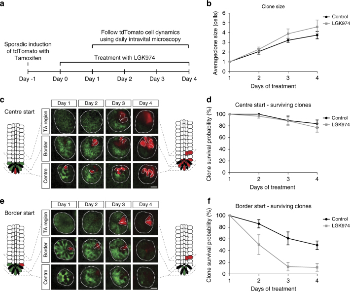
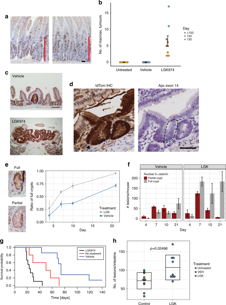
Many epithelial stem cell populations follow a pattern of stochastic stem cell divisions called 'neutral drift'. It is hypothesised that neutral competition between stem cells protects against the acquisition of deleterious mutations. Here we use a Porcupine inhibitor to reduce Wnt secretion at a dose where intestinal homoeostasis is maintained despite a reduction of Lgr5+ stem cells. Functionally, there is a marked acceleration in monoclonal conversion, so that crypts become rapidly derived from a single stem cell. Stem cells located further from the base are lost and the pool of competing stem cells is reduced. We tested whether this loss of stem cell competition would modify tumorigenesis. Reduction of Wnt ligand secretion accelerates fixation of Apc-deficient cells within the crypt leading to accelerated tumorigenesis. Therefore, ligand-based Wnt signalling influences the number of stem cells, fixation speed of Apc mutations and the speed and likelihood of adenoma formation.


Conflict of interest statement

M.E.M. is an employee of Novartis Inc. The remaining authors declare no competing interests.

Figures

Fig. 1
Fig. 1
Homoeostasis is unperturbed after LGK974 treatment. a C57BL/6 mice were treated with LGK974 for 4.5 days. The small intestine showed no toxicity and no difference in proliferation (BrdU). Treatment of LGK974 leads to downregulation of the intestinal stem cell genes Lgr5 and Olfm4 as confirmed by RNA in situ hybridisation. Note only a few cells at the bottom of the crypt still express Olfm4 after LGK974 treatment (N = 3 for both groups). b Quantification of BrdU+ cells/half-crypt (at least 30 crypts per mouse were analysed). Each dot represents the average per mouse, red bar = mean per group. Vehicle (VEH) N = 3, LGK974 (LGK) N = 4. c qRT-PCR confirms downregulation of several stem cell genes, whereas expression of Bmi1 is unchanged. Each dot represents single mouse sample, black bar indicates mean per group, N = 3 per group. d Uninduced Catnblox(ex3)/lox(ex3) are hypomorphs with about 50% reduced expression of β-catenin (Ctnnb1), as confirmed by qRT-PCR (N = 3). e Reduced expression of β-catenin results in hyper-sensitivity to LGK974 and loss of the (small) intestinal crypts within 10 days (mean survival), N = 8. Scale bar = 50 µm
Fig. 2
Fig. 2
Stem cell replacement rate is accelerated after LGK974 treatment. Mice were induced with 0.15 mg tamoxifen to induce tdTomfl recombination in few Lgr5-CreER-EGFP+ cells. a Mosaic expression of the Lgr5-eGFP (green) and the recombined tdTomfl+ cells (red). Nuclei were stained with DAPI (blue). Representative pictures at day 10, note the loss of GFP expression and clones are fully labelled by tdTom+ cells in Porcupine inhibitor-treated mice (LGK974). Scale bar = 100 um. b Clone size was counted in 'eighths', at time indicated after induction. At least 200 clones per mouse were counted, vehicle (VEH) N = 3, 3, 4, 4, 3, 3 and LGK974 (LGK) N = 2, 4, 3, 5, 3, 3 for each timepoint, respectively. Heatmap shows all counted clones per timepoint/group. Note that LGK974 has an increased clone size at day 4 and the mean clone size from day 7 is almost at its maximum. Graph shows the mean clone size (c) or the number of fully fixed crypts (d) at different time points as shown in b. Error bars, s.e.m. e Number of tdTom+ clones per field. The number of crypts with at least one tdTom+ cell were counted per field, ≥19 images per mouse, error bars = s.e.m. Note the similar number of clones at day 4 but the greater reduction in clones after LGK974 treatment at day 10. Vehicle (VEH) N = 3, 3, 4, 4, 3, 3 and LGK974 (LGK) N = 2, 4, 3, 5, 3, 3 for each timepoint respectively
Fig. 3
Fig. 3
In vivo live imaging shows specific loss of border stem cells after LGK974 treatment. a Graphical representation of the experimental setup. One day before LGK974 treatment, Lgr5CreERtdTomfl mice were injected with 0.05 mg tamoxifen IP to induce recombination in single cells. Mice (N = 4) were treated with LGK974 and daily intravital imaging was performed starting 1 day after first LGK974 treatment and compared to control mice (N = 5). b Graph shows mean clone size (43 crypts, control; 56 crypts, LGK974 on day 1) of surviving clones (clones with at least one cell in centre or border) over time within the centre and border. Note that cells in the transit amplifying (TA) cell region are not counted. c, e Intravital images of the same crypt on days 1–4 following a clone starting in the centre (c) and a clone starting in the border (e). d, f Graphs show the percentage of clones that still have at least one cell in the centre or border starting from centre cell (17 crypts, control; 36 crypts, LGK974) (d) or from border cell (26 crypts, control; 20 crypts, LGK974) (f). Error bars = s.e.m. Scale bar, 20 µm
Fig. 4
Fig. 4
Reduced Wnt ligand secretion does not affect Apc-deficient cell growth but accelerates tumorigenesis. aVilCreERApcfl/fl mice were induced and treated with LGK974 starting the following day. Mice were sampled at day 4 post induction, and proliferation assessed by BrdU staining. Scale bar = 50 µm. bLgr5CreERApcfl/fl mice induced with 0.15 mg tamoxifen and treated with LGK974/vehicle 1 day p.i. The number of macroscopic adenomas was scored when mice showed signs of intestinal adenomas or at 100/130 day timepoint. Each dot represents one mouse, untreated N = 5, vehicle N = 7 and LGK974 N = 9, black dot = mean, error bars = s.e.m., Mann–Whitney U test: untreated vs. LGK974 p = 0.002639, vehicle vs. LGK974 p = 0.0006014. c Immunohistochemistry for β-catenin showed only small microadenomas in the vehicle group, in contrast to adenomas found after LGK974 treatment. Scale bar = 100 µm. d Example image of Lgr5CreERApcfl/fltdTomfl/+ mouse 10 days post induction (3 mg, LGK974 treatment). Immunohistochemistry for tdTomato (RFP) shows fully labelled crypt. Arrow marks unlabelled cell (probably Paneth cell), suggesting a newly labelled crypt. RNA in situ for Apc exon 14 shows that only half of the crypt has recombined (dashed area). Scale bar = 50 µm. eLgr5CreER Apcfl/fl mice were induced with 3 mg tamoxifen and treated with LGK974/vehicle starting at day 1 p.i. Crypts were scored based on immunohistochemistry for β-catenin and categorised into partial and full crypts. The ratio of full crypts is in relation to the sum of full and partial crypts. f Scoring of partial and full crypts at different timpoints in absolute numbers. N = 3 mice for each timepoint and each group, error bars = s.e.m. gLgr5CreER Apcfl/fl mice induced with 3 mg tamoxifen were treated with LGK974 or vehicle starting at day 1 p.i. Mice were sampled when signs of intestinal tumour burden were apparent. Untreated N = 5, vehicle (VEH) N = 7, LGK974 N = 9 mice, log-rank test: vehicle vs. LGK974 p = 0.00019, untreated vs. LGK974 p = 0.0222. h Number of adenomas scored on histological sections. Untreated N = 5, vehicle (VEH) N = 6, LGK974 N = 9 mice. Each dot represents number of lesions per mouse; box indicates mean ± standard deviation. Mann–Whitney U test for LGK974 vs. control mice (untreated and vehicle) p = 0.02496

References

    1. Ritsma L, et al. Intestinal crypt homeostasis revealed at single-stem-cell level by in vivo live imaging. Nature. 2014;507:362–365. doi: 10.1038/nature12972. - DOI - PMC - PubMed
    1. Kozar, S. et al. Continuous clonal labeling reveals small numbers of functional stem cells in intestinal crypts and adenomas. Cell Stem Cell13, 626–633 (2013).. - PubMed
    1. de Lau W, et al. Lgr5 homologues associate with Wnt receptors and mediate R-spondin signalling. Nature. 2011;476:293–297. doi: 10.1038/nature10337. - DOI - PubMed
    1. Farin, H. F., Van Es, J. H. & Clevers, H. Redundant sources of Wnt regulate intestinal stem cells and promote formation of Paneth cells. Gastroenterology. doi: https://doi.org/10.1053/j.gastro.2012.08.031 (2012). - PubMed
    1. Tian H, et al. A reserve stem cell population in small intestine renders Lgr5-positive cells dispensable. Nature. 2011;478:255–259. doi: 10.1038/nature10408. - DOI - PMC - PubMed

Publication types

MeSH terms