. 2018 Mar 19;8(1):4853.
doi: 10.1038/s41598-018-23258-w.

Vitamin D alleviates lead induced renal and testicular injuries by immunomodulatory and antioxidant mechanisms in rats

Affiliations

Vitamin D alleviates lead induced renal and testicular injuries by immunomodulatory and antioxidant mechanisms in rats

Mohammad A BaSalamah et al. Sci Rep. .

Abstract

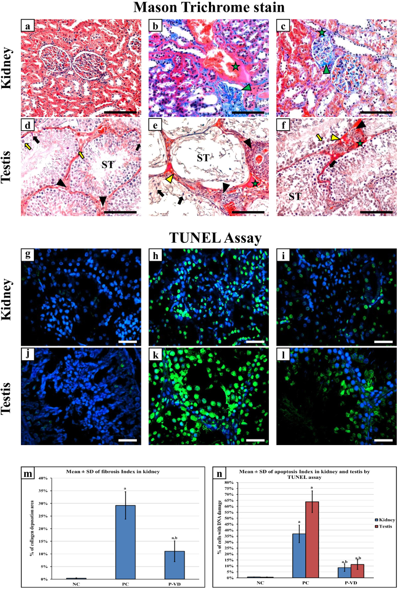
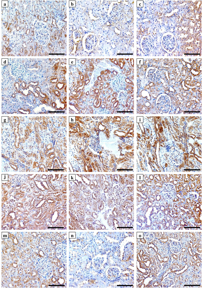
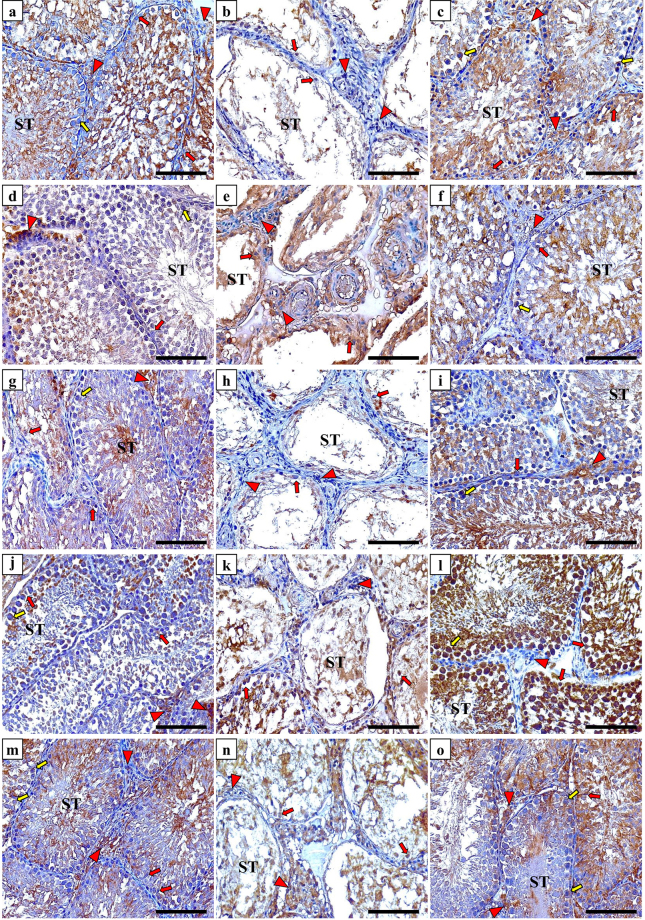
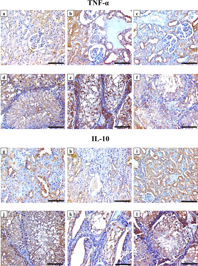
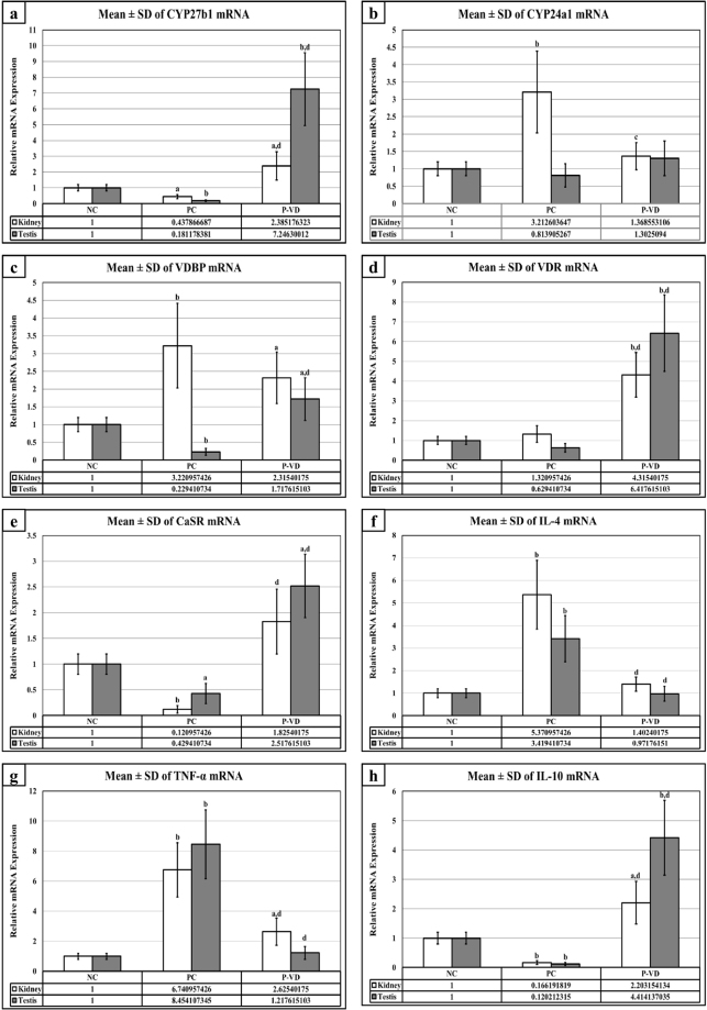
This study measured the effects of vitamin D (VD) supplementation on the underlying molecular pathways involved in renal and testicular damage induced by lead (Pb) toxicity. Thirty two adult male Wistar rats were divided equally into four groups that were treated individually or simultaneously, except the negative control, for four weeks with lead acetate in drinking water (1,000 mg/L) and/or intramuscular VD (1,000 IU/kg; 3 days/week). Pb toxicity markedly reduced serum VD and Ca2+, induced substantial renal and testicular injuries with concomitant significant alterations in the expression of VD metabolising enzymes, its receptor and binding protein, and the calcium sensing receptor. Pb also significantly promoted lipid peroxidation and pro-inflammatory cytokines (IL-4 and TNF-α) in the organs of interest concomitantly with declines in several anti-oxidative markers (glutathione, glutathione peroxidase and catalase) and the anti-inflammatory cytokine, IL-10. The co-administration of VD with Pb markedly mitigated renal and testicular injuries compared with positive controls. This was associated with restoration of the expression of VD related molecules, promotion of anti-oxidative and anti-inflammatory markers, but tissue Pb concentrations were unaffected. In conclusion, this report is the first to reveal potential protective effects for VD against Pb-induced renal and testicular injuries via anti-inflammatory and anti-oxidative mechanisms.


Conflict of interest statement

The authors declare no competing interests.

Figures

Figure 1
Figure 1
Histopathological features by Mason’s trichrome stain of renal and testicular tissues of the NC (a & d), PC (b & e), and P-VD (c & f) groups (40× objective, scale bar = 8 µm; green star = interstitial necrosis; green arrow head = collagen type I deposition in glomerulus; yellow arrow head = hyaline droplets; ST = seminiferous tubule; black arrow = Sertoli cell; black arrow head = Leydig cell and yellow arrow = spermatogonia). Cellular DNA damage was detected by TUNEL assay (Green fluorescent dye) in kidney and testis from the NC (a & g), PC (b & h), and P-VD (c & i) groups (40× objective; scale bar = 10 µm; positive nuclei are stained in green). Additionally, the renal fibrosis index (m) and renal and testicular apoptosis/necrosis index (n) are shown as bar graphs for the study groups. (a = P < 0.05 compared with NC group and b = P < 0.05 compared with PC group).
Figure 2
Figure 2
Immunohistochemical expression of Cyp27b1 enzyme (upper row), Cyp24a1 enzyme (2nd row from top), VDBP (3rd row from top), VDR (4th row from top) and CaSR (bottom row) in kidney sections from the NC (left column), PC (middle column) and P-VD (right column) groups (40× objective, scale bar = 8 µm).
Figure 3
Figure 3
Immunohistochemical expression of Cyp27b1 enzyme (upper row), Cyp24a1 enzyme (2nd row from top), VDBP (3rd row from top), VDR (4th row from top) and CaSR (bottom row) in testicular sections from the NC (left column), PC (middle column) and P-VD (right column) groups (40× objective; scale bar = 8 µm; ST = seminiferous tubule; red arrow = Sertoli cell; red arrow head = Leydig cell and yellow arrow = spermatogonia).
Figure 4
Figure 4
Immunohistochemical expression of TNF-α and IL-10 in renal (1st and 3rd rows from top) and testicular (2nd and 4th rows from top) sections from the NC (left column), PC (middle column) and P-VD (right column) groups (40× objective, scale bar = 8 µm).
Figure 5
Figure 5
Mean ± SD of messenger RNA relative expression of (a) Cyp27b1, (b) Cyp24a1, (c) VDBP, (d) VDR, (e) CaSR, (f) IL-4, (g) TNF-α and (h) IL-10 in the different study groups. (a = P < 0.05 compared with the NC group; b = P < 0.01 compared with the NC group; c = P < 0.05 compared with the PC group and d = P < 0.01 compared with the PC group).

Similar articles

Cited by

References

    1. Caito, S., Lopes, A., Paoliello, M. M. B., & Aschner, M. Toxicology of Lead and Its Damage to Mammalian Organs. 2017/07/22 ed. Met Ions Life Sci. Vol. 17 (2017). - PubMed
    1. Ericson B, et al. The Global Burden of Lead Toxicity Attributable to Informal Used Lead-Acid Battery Sites. Ann Glob Health. 2016;82:686–699. doi: 10.1016/j.aogh.2016.10.015. - DOI - PubMed
    1. Haryanto B. Lead exposure from battery recycling in Indonesia. Rev Environ Health. 2016;31:13–6. doi: 10.1515/reveh-2015-0036. - DOI - PubMed
    1. World Health Organization (WHO). Lead poisoning and health. 2016; Available from: http://www.who.int/mediacentre/factsheets/fs379/en/ [Accessed 2017 28/09].
    1. Gandhi J, et al. Impaired hypothalamic-pituitary-testicular axis activity, spermatogenesis, and sperm function promote infertility in males with lead poisoning. Zygote. 2017;25:103–110. doi: 10.1017/S0967199417000028. - DOI - PubMed

MeSH terms