. 2018 Apr;20(4):393-399.
doi: 10.1038/s41556-018-0068-5. Epub 2018 Mar 19.

EFF-1 fusogen promotes phagosome sealing during cell process clearance in Caenorhabditis elegans

Affiliations

EFF-1 fusogen promotes phagosome sealing during cell process clearance in Caenorhabditis elegans

Piya Ghose et al. Nat Cell Biol. 2018 Apr.

Abstract

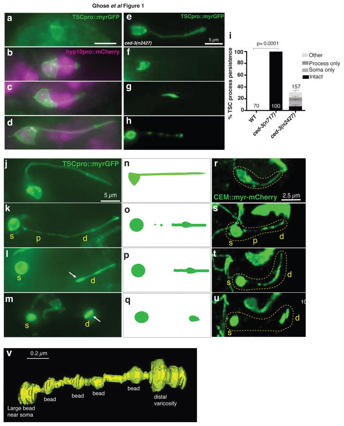
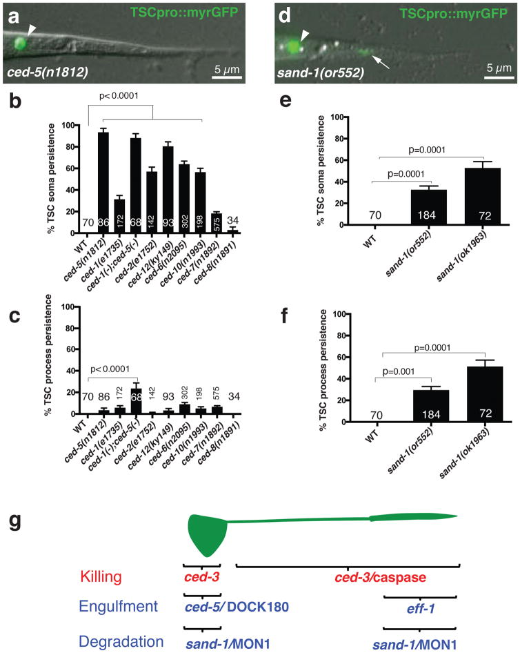
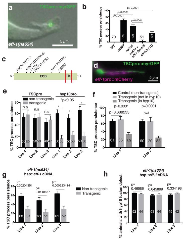
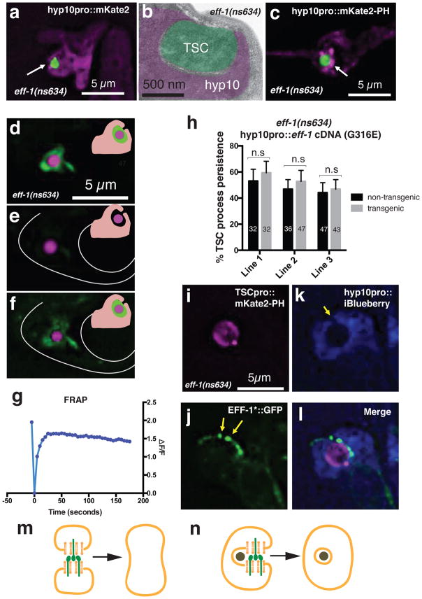
Phagocytosis of dying cells is critical in development and immunity1-3. Although proteins for recognition and engulfment of cellular debris following cell death are known4,5, proteins that directly mediate phagosome sealing are uncharacterized. Furthermore, whether all phagocytic targets are cleared using the same machinery is unclear. Degeneration of morphologically complex cells, such as neurons, glia and melanocytes, produces phagocytic targets of various shapes and sizes located in different microenvironments6,7. Thus, such cells offer unique settings to explore engulfment programme mechanisms and specificity. Here, we report that dismantling and clearance of a morphologically complex Caenorhabditis elegans epithelial cell requires separate cell soma, proximal and distal process programmes. Similar compartment-specific events govern the elimination of a C. elegans neuron. Although canonical engulfment proteins drive cell soma clearance, these are not required for process removal. We find that EFF-1, a protein previously implicated in cell-cell fusion 8 , specifically promotes distal process phagocytosis. EFF-1 localizes to phagocyte pseudopod tips and acts exoplasmically to drive phagosome sealing. eff-1 mutations result in phagocytosis arrest with unsealed phagosomes. Our studies suggest universal mechanisms for dismantling morphologically complex cells and uncover a phagosome-sealing component that promotes cell process clearance.


Figures

Figure 1
Figure 1. The tail-spike cell (TSC) and CEM neurons undergo a similar degeneration sequence
(a–d) TSC in comma stage, 1.5 fold, 2-fold, and 3-fold embryos, respectively and its association with hyp10. n=5 biologically independent animals each with similar results. Scale bar: 5 μm. (e–h) ced-3(n2427) mutants exhibit an intact cell, persistent soma only, persistent process only, or intermediate degeneration (other), respectively. n=10 biologically independent animals each with similar results. Scale bars: 5 μm. (i) TSC persistence in ced-3 mutants. Data are mean +/− s.e.m. Statistics: two-tailed unpaired t-test. Individual p values: see Supplementary Table 2. Numbers inside/outside bars, total animals scored per genotype. Data are from 3 independent scoring experiments. n=sample sizes for statistics are as follows: WT:70, ced-3(n717):100, ced-3(n2427): 157 (other: 42, process only: 31, soma only:47, intact: 37. (j) TSC in 3-fold embryo. (k) TSC soma rounding and proximal process beading. (l,m) distal process retraction. n=10 biologically independent animals each. Scale bar: 5 μm. (r–u) Stills from movies showing CEM death. Scale bar: 2.5 μm. Time: hours:minutes post-fertilization, 07:40 (r), 08:05 (s), 08:15 (t), 08:30 (u). (n–q) schematics for j–m and r–u. n=6 biologically independent animals each. Yellow text: s, soma; p, proximal process; d, distal process. White arrowhead, distal varicosity. (v) 3D reconstruction of a degenerating TSC in an eff-1(ns634) animal. TSC death stages in this mutant are similar to WT (Main text, Supplementary Movie 5). Reconstruction based on Supplementary Movie 2. Scale bar: 0.2 μm. Statistics source data are provided in Supplementary Table 2.
Figure 2
Figure 2. Canonical engulfment genes are not required for TSC process clearance
(a) Unengulfed soma in ced-5(n1812) L1 animal. n=86 biologically independent animals with similar results. (b) TSC soma clearance defects. (c) TSC process clearance defects. n=sample sizes for statistics are as follows, with n referring to number of biologically independent animals.WT: n=70, ced-5(n1812): n=86, ced-1(e1735): n=172, ced-1(-);ced-5(-):n=68, ced-2(e1752):n=142, ced-6(n2095):n=302, ced-10(n1993):n=198, ced-7(n1892):n=575, ced-8(n1981): n=34. (d) Undegraded soma and process in sand-1(or552) L1 animal. n=184 biologically independent animals with similar results. (e,f). TSC soma, process degradation defects in sand-1 mutants, respectively. n=sample sizes for statistics are as follows, with n referring to number of biologically independent animals.WT:n=70, sand-1(or552):n=184), sand-1(ok1963):n=72 (g). Genes promoting TSC regional killing and clearance. Arrowhead, soma; arrow, process. Data are mean +/− s.e.m. Statistics: two-tailed unpaired t-test. Individual p values: see Supplementary Table 2. Numbers inside bars, total animals scored per genotype. 3 independent scoring experiments were done. Scale bar: 5 μm. Statistics source data are provided in Supplementary Table 2.
Figure 3
Figure 3. EFF-1 fusogen mediates distal process clearance
(a) L1 eff-1(ns634) mutant. n=129 biologically independent animals with similar results. (b) Process clearance defects in indicated genotypes. n=sample sizes for statistics are as follows with n referring to number of biologically independent animals.WT: n=70, ns627: n=100, ns634:n=129, ns634+ fosmid: n=51, eff-1(hy21):n=108. (c) EFF-1 structure and mutation sites. ECD, extracellular domain; TM, transmembrane domain. hy41 was previously identified. (d) ced-3(n717) mutant showing hyp10 eff-1 expression. n=4 biologically independent animals with similar results. (e) eff-1 mutant cell-specific rescue. n=sample sizes for statistics are as follows with n referring to number of biologically independent animals: TSC Line 1: n=50 (non transgenic), n=50 (transgenic); TSC Line 2: n=50 (non transgenic), n=50 (transgenic); TSC Line 3: n=49 (non transgenic), n=47 (transgenic); hyp10 Lines 1–3 each: n=50 (non transgenic), n=50 (transgenic) (f) Mosaic analysis showing EFF-1 function in hyp10 for TSC distal process clearance. n=sample sizes for statistics are as follows, Line 1: Control: 61, Transgenic (not in hyp10): n=33, Transgenic (in hyp10): n=89; Line 2: Control: n=60, Transgenic (not in hyp10): n=20, Transgenic (in hyp10): n=83. (g) Rescue of eff-1 mutant TSC clearance defect with heat-shock promoter::eff-1 cDNA following heat exposure. n=sample sizes for statistics are as follows, Line 1: n=60 (non-transgenic), n=51 (transgenic); Line 2: n=63 (non-transgenic), n=51 (transgenic); Line 3: n=61 (non-transgenic), n=52 (transgenic). (h) Same as g, except rescue of fusion examined. n=sample sizes for statistics are as follows, Line 1: n=52 (non-transgenic), n=44 (transgenic); Line 2: n=53 (non-transgenic), n=44 (transgenic); Line 3: n=45 (non-transgenic), n=42 (transgenic). Data are mean +/− s.e.m. Statistics: two-tailed unpaired t-test. n.s., non significant (p>0.05). Individual p values: see Supplementary Table 2. Numbers inside bars, total animals scored per genotype. For rescue experiments, 2–3 independent transgenic lines were scored. For other experiments, 3 independent scoring experiments were done. For heat-shock rescue, scoring was done 2 hours after heat-shock. Scale bars: 5 μm. Statistics source data are provided in Supplementary Table 2.
Figure 4
Figure 4. EFF-1 promotes phagosome sealing
(a) TSC distal process incompletely engulfed by hyp10 cell. Arrow, phagosome opening. n=15 biologically independent animals with similar results. (b). Electron micrograph of eff-1(ns634) L1 showing open phagosome. Area not pseudocolored, cuticle. n=1 animal. (c) Enrichment of open phagosome marker (arrow) around TSC fragment. n=10 biologically independent animals with similar results. (d–f) FRAP experiment in eff-1(ns634); cup-2(ar506); ssGFP: green, secreted GFP; magenta, TSC; inset, secreted GFP (green) surrounding TSC in phagosome space. (d) before, (e) immediately after, (f) 30 minutes after photobleaching. n=11 biologically independent animals with similar results. (g) Quantification of a different FRAP experiment. n=19 biologically independent animals with similar results. (h) EFF-1(G316E) cannot rescue eff-1(ns634) distal fragment clearance defect. n=sample sizes for statistics are as follows, Line 1: n=32 (non-transgenic), n=32 (transgenic); Line 2: n=36 (non-transgenic), n=47 (transgenic); Line 3: n=47 (non-transgenic), n=43 (transgenic). (i–l) Co-localization of EFF-1 (arrowheads) and phagosome opening (arrow). n=4 biologically independent animals with similar results. (m,n) Models of EFF-1 (green) function in cell-cell fusion and phagosome sealing, respectively. Data are mean +/− s.e.m. Statistics: two-tailed unpaired t-test. Individual p values: see Supplementary Table 2. n.s., not significant (p>0.05). Numbers inside bars, total animals scored per genotype. 3 independent transgenic lines were scored for rescue experiment. FRAP experiments were repeated 38 times. Scale bars: 5 μm.

Similar articles

Cited by

References

    1. Lekstrom-Himes JA, Gallin JI. Immunodeficiency diseases caused by defects in phagocytes. The New England journal of medicine. 2000;343:1703–1714. doi: 10.1056/NEJM200012073432307. - DOI - PubMed
    1. Levin R, Grinstein S, Canton J. The life cycle of phagosomes: formation, maturation, and resolution. Immunological reviews. 2016;273:156–179. doi: 10.1111/imr.12439. - DOI - PubMed
    1. Mallat M, Marin-Teva JL, Cheret C. Phagocytosis in the developing CNS: more than clearing the corpses. Current opinion in neurobiology. 2005;15:101–107. doi: 10.1016/j.conb.2005.01.006. - DOI - PubMed
    1. Hochreiter-Hufford A, Ravichandran KS. Clearing the dead: apoptotic cell sensing, recognition, engulfment, and digestion. Cold Spring Harbor perspectives in biology. 2013;5:a008748. doi: 10.1101/cshperspect.a008748. - DOI - PMC - PubMed
    1. Reddien PW, Horvitz HR. The engulfment process of programmed cell death in caenorhabditis elegans. Annual review of cell and developmental biology. 2004;20:193–221. doi: 10.1146/annurev.cellbio.20.022003.114619. - DOI - PubMed

Publication types

MeSH terms

Substances