Review
. 2018 Jun;19(6):385-397.
doi: 10.1038/s41576-018-0001-6.

Molecular mechanisms driving transcriptional stress responses

Affiliations
Review

Molecular mechanisms driving transcriptional stress responses

Anniina Vihervaara et al. Nat Rev Genet. 2018 Jun.

Abstract

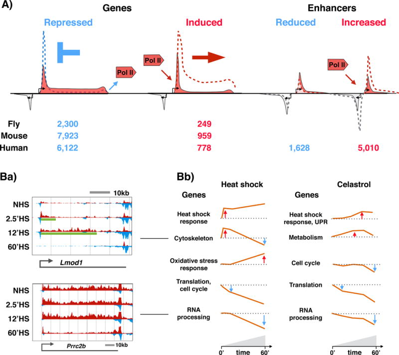
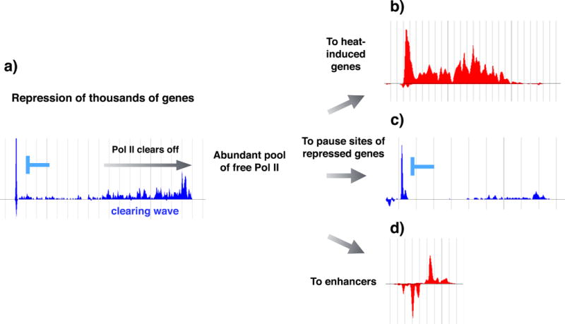
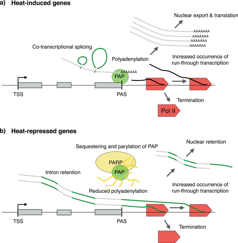
Proteotoxic stress, that is, stress caused by protein misfolding and aggregation, triggers the rapid and global reprogramming of transcription at genes and enhancers. Genome-wide assays that track transcriptionally engaged RNA polymerase II (Pol II) at nucleotide resolution have provided key insights into the underlying molecular mechanisms that regulate transcriptional responses to stress. In addition, recent kinetic analyses of transcriptional control under heat stress have shown how cells 'prewire' and rapidly execute genome-wide changes in transcription while concurrently becoming poised for recovery. The regulation of Pol II at genes and enhancers in response to heat stress is coupled to chromatin modification and compartmentalization, as well as to co-transcriptional RNA processing. These mechanistic features seem to apply broadly to other coordinated genome-regulatory responses.


Conflict of interest statement

Competing interests statement

The authors declare no competing interests.

Figures

Figure 1
Figure 1. Sensing, communicating and transcriptionally responding to protein-damaging stress
Stress conditions in different compartments of the cell need distinct trajectories to communicate with the nucleus to provoke a transcriptional change. a) In the cytosol, heat stress causes heat shock proteins (HSPs) to release heat shock factor 1 (HSF1), which then can trimerize, bind to its target DNA elements and transactivate genes that encode chaperone machineries and polyubiquitin (reviewed in,). b) Oxidative stress inactivates prolyl hydroxylase domain-containing proteins (PHDs), which reduces the degradation of hypoxia-induced factor 1α (HIF1α), allowing HIF1α to dimerize with HIF1β and activate transcriptional programmes for induced angiogenesis and oxygen supply (reviewed in). c) In the endoplasmic reticulum (ER), protein misfolding triggers the unfolded protein response (UPRER), which communicates from the ER lumen to the cytosol via the membrane-embedded proteins inositol-requiring protein 1 (IRE1), protein kinase R (PKR)-like ER kinase (PERK), and cyclic AMP-dependent transcription factor ATF6 (,,– and references therein). Each of these three pathways leads to the generation of distinct transactivators (by activated splicing and translation of X-box binding protein 1 (XBP1), by translation control of ATF4 or by proteolytic processing of ATF6, respectively). d) In the mitochondria, misfolded proteins are first processed by protease complexes (grey sliced circle). The peptides move to the cytosol via ATP-binding cassette (ABC) transporters (white barrels), subsequently activating the mitochondrial UPR (UPRMT)-responsive transactivators ATF5 and DNA damage-inducible transcript 3 protein (DDIT3; also known as CHOP),,. In a and b, heat and hypoxia lead to a rapid activation of constitutively expressed transactivators that are maintained inactive by the complexes that sense misfolded proteins or levels of oxygen, respectively (reviewed in,,). In c and d, compartment-restricted stresses are communicated over membrane barriers, which likely delays the transcriptional responses.
Figure 2
Figure 2. Heat shock response triggers transcriptional repro o ramming of genes and enhancers across the genome
A Schematic representation of RNA polymerase II (Pol II) distribution at heat-responsive genes and enhancers. Heat stress provokes repression of thousands and induction of hundreds of genes in flies, mice and humans. In mammals, the repression is mediated by inhibiting the release of paused Pol II into elongation, causing accumulation of Pol II at the pause site (blue dashed line. In flies (nuoe.t shown), the heat repression causes decreased Pol II density across the gene. From fly to human, the rapid activation of genes is triggered by releasing promoter-proximal paused Pol II into elongation (red dashed line). Concurrent with reprogramming of genes, the enhancer landscape is re-profiled in eat-stressed cells, provoking educed or increased Pol II density at thousands of enhancers. Gene transcription is depicted with red; divergent transcription with light grey. At enhancers, both strands (indicated with red and grey) produce short and unstable transcripts. B) Temporal patterns of heat-responsive or celastrol-responsive gene regulation. Ba) Browser-shot examples of rapidly, but transiently, expressed Lmod1 (upper panel) and late repressed Prrc2b (lower panel) genes in wild-type mouse cells under normal conditions (0) and under heat stress of different duration (2.5, 12 and 60 minutes). The transcriptional profile of the plus (red) and minus (blue) strand is depicted. The green line under the transcription profiles (upper panel) indicates the distance that the heat-induced Pol II complexes have travelled at the indicated time point. Bb) Five major temporal patterns appear in heat-shocked (left panels) or celastrol-treated (right panels) cells. The orange line recapitulates the average fold change with respect to the basal transcription level (dotted line) in each category. The main gene ontology terms associated, with each group are indicated to the left of the profile. UPR, unfolded protein response. Panels in Ba and Bb are adapted with permission from and .
Figure 3
Figure 3. Promoter opening, establishment of directionality and rapid release of paused RNA polymerase II
Before heat shock, GAGA-associated Factor (GAF) and nucleosome remodelling factor (NURF) create nucleosome-free promoters at heat-induced genes in Drosophila melanogaster, where strong promoter elements direct the pre-initiation initiation complex (PIC), and hence RNA polymerase II (Pol II), to the coding strand (part a). In mammals, replication protein A (RPA) and the FACT (facilitates chromatin transcription) complex contribute to promoter opening (part b). Preferential binding of the PIC to the core promoter of the coding strand creates directionality by orienting Pol II towards the gene. The highly paused and highly directional genes are rapidly activated by releasing paused Pol II into productive elongation in flies (part c) and mammals (part d). The pause-release is triggered by heat-activated transactivators, such as heat shock factor 1 (HSF1) (parts c and d) and possibly serum response factor (SRF) (part d), through (direct or indirect) recruitment of positive transcription elongation factor b (P-TEFb) and chromatin modifiers. Nucleosome remodelling by FACT and acetyltransferases allows efficient Pol II progression along the gene. Sumoylation (S) and poly(ADP-ribose) polymerase (PARP1)-mediated parylation can modify transcriptional regulators and facilitate chromatin compartmentalization. The arrows indicate recruitment of Pol II (grey), transactivators HSF1 and SRF (red) and P-TEFb (purple). The shaded grey areas demonstrate Pol II density. Ac, acetylation; DSIF, DRB sensitivity-inducing inducing factor; NELF, negative elongation factor; P, phosphorylation.
Figure 4
Figure 4. Redistribution of transcription machinery upon heat shock
Inhibition of pause-release causes RNA polymerase II (Pol II) to clear off from thousands of gene bodies (part a). In mammals, where genes are generally long, the clearing of Pol II causes an abundant pool of unengaged Pol II in the cell. The increased availability of Pol II can be utilized for rapid filling of the pause sites at heat-induced genes where the rate of pause-release profoundly increases (part b), as well as the filling of the available pause sites at heat-repressed genes where inhibition of pause-release causes high promoter-proximal pause signal (part c). The increased availability of Pol II is likely to be also contributing to the tuning up of the enhancer repertoire, as an increased number and transcription of enhancers are detected upon heat11 and celastrol28 stresses (part d). The Pol II profile at these example genes and enhancer is obtained from the raw data presented in ref., where human K562 cells were subjected to a 30-minute heat stress at 42 °C. Transcriptionally repressed genes (parts a and c) are indicated in blue, and a transcriptionally induced gene (part b) and enhancer (part d) are depicted in red.
Figure 5
Figure 5. Co-transcriptional processing in heat-stressed cells
Transcripts of heat-induced genes are co-transcriptionally spliced, efficiently polyadenylated and exported to the cytosol (part a). By contrast, transcripts of a number of uninduced genes retain introns, show reduced polyadenylation and increased readthrough transcription, and are retained in the nucleus during heat exposure (part b). Retention of intron-containing transcripts could be a mechanism to store pre-mRNA until the stress is ameliorated and cellular processes are restored. PAP, poly(A) polymerase; PARP1, poly(ADP-ribose) polymerase 1; PAS, polyadenylation and cleavage site; Pol II, RNA polymerase II; TSS, transcription start site.

References

    1. Richter K, Haslbeck M, Buchner J. The heat shock response: life on the verge of death. Mol Cell. 2010;40:253–266. - PubMed
    1. Toivola DM, Strnad P, Habtezion A, Omary MB. Intermediate filaments take the heat as stress proteins. Trends Cell Biol. 2010;20:79–91. - PMC - PubMed
    1. Walter P, Ron D. The unfolded protein response: from stress pathway to homeostatic regulation. Science. 2011;334:1081–1086. - PubMed
    1. Quirós PM, Langer T, López-Otín C. New roles for mitochondrial proteases in health, ageing and disease. Nat Rev Mol Cell Biol. 2015;16:345–359. - PubMed
    1. Majmundar AJ, Wong WJ, Simon MC. Hypoxia-inducible factors and the response to hypoxic stress. Mol Cell. 2010;40:294–309. - PMC - PubMed

Publication types

LinkOut - more resources