. 2018 Mar 5:9:183.
doi: 10.3389/fphar.2018.00183. eCollection 2018.

Curcumin Prevents Acute Neuroinflammation and Long-Term Memory Impairment Induced by Systemic Lipopolysaccharide in Mice

Affiliations

Curcumin Prevents Acute Neuroinflammation and Long-Term Memory Impairment Induced by Systemic Lipopolysaccharide in Mice

Vincenzo Sorrenti et al. Front Pharmacol. .

Abstract

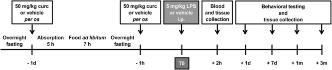
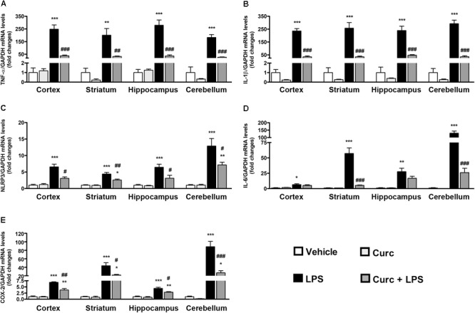
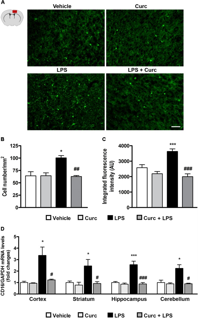
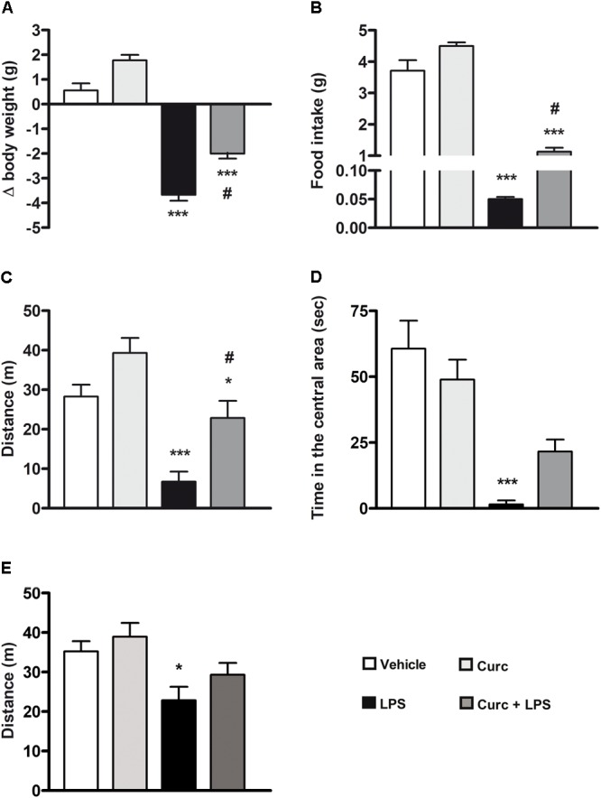
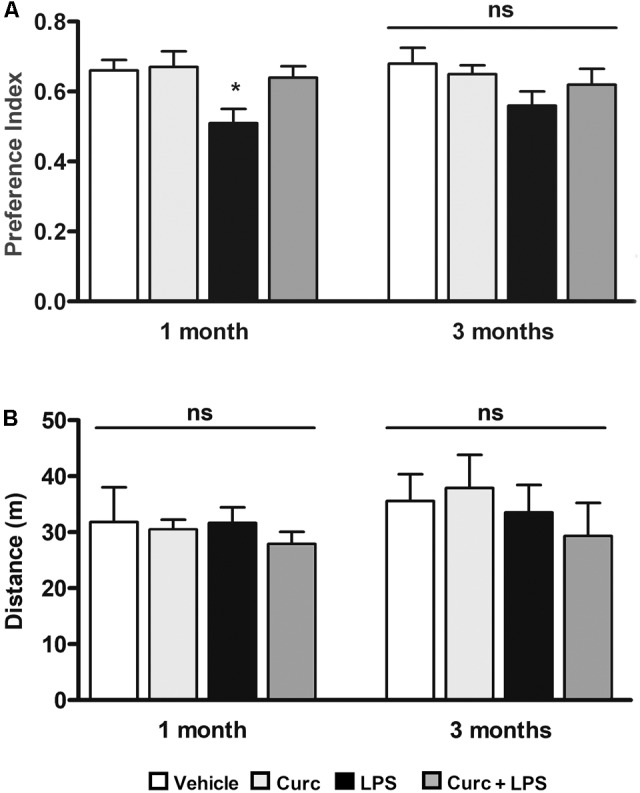
Systemic lipopolysaccharide (LPS) induces an acute inflammatory response in the central nervous system (CNS) ("neuroinflammation") characterized by altered functions of microglial cells, the major resident immune cells of the CNS, and an increased inflammatory profile that can result in long-term neuronal cell damage and severe behavioral and cognitive consequences. Curcumin, a natural compound, exerts CNS anti-inflammatory and neuroprotective functions mainly after chronic treatment. However, its effect after acute treatment has not been well investigated. In the present study, we provide evidence that 50 mg/kg of curcumin, orally administered for 2 consecutive days before a single intraperitoneal injection of a high dose of LPS (5 mg/kg) in young adult mice prevents the CNS immune response. Curcumin, able to enter brain tissue in biologically relevant concentrations, reduced acute and transient microglia activation, pro-inflammatory mediator production, and the behavioral symptoms of sickness. In addition, short-term treatment with curcumin, administered at the time of LPS challenge, anticipated the recovery from memory impairments observed 1 month after the inflammatory stimulus, when mice had completely recovered from the acute neuroinflammation. Together, these results suggest that the preventive effect of curcumin in inhibiting the acute effects of neuroinflammation could be of value in reducing the long-term consequences of brain inflammation, including cognitive deficits such as memory dysfunction.

Keywords: curcumin; lipopolysaccharide; memory impairment; microglia; neuroinflammation; pro-inflammatory cytokines; sickness behavior.


Figures

FIGURE 1
FIGURE 1
Schematic representation of the experimental design of this study. Mice treated for 2 consecutive days with curcumin (curc; 50 mg/kg) received a single intraperitoneal (i.p.) injection of LPS (5 mg/kg). Control animals received per os the same volume of vehicle following the same time schedule and were then injected i.p. with saline. At different times after LPS administration, behavioral tests were performed and blood samples and brain tissues were collected for analysis.
FIGURE 2
FIGURE 2
Curcumin inhibits the acute brain pro-inflammatory response to peripheral LPS administration. Mice treated for 2 consecutive days with curcumin (curc; 50 mg/kg) received a single i.p. injection of LPS (5 mg/kg). Two hours following LPS or saline injection, mice were sacrificed and cerebral cortex, striatum, hippocampus, and cerebellum were extracted and prepared as described in Section “Materials and Methods.” (A) Analysis of TNF-α mRNA expression levels was conducted via real-time PCR. Results are expressed as fold-increase with respect to control (vehicle only). Data are means ± SEM (n = 4 mice/group). Data were analyzed by one-way ANOVA [F(15,48) = 32.89, p < 0.0001] followed by Bonferroni’s multiple comparison test. ∗∗∗p < 0.001 vs. control group; ##p < 0.01 and ###p < 0.001 vs. LPS-stimulated group. (B) Analysis of IL-1β mRNA expression levels was conducted via real-time PCR. Results are expressed as fold-increase with respect to control (vehicle only). Data are means ± SEM (n = 4 mice/group). Data were analyzed by one-way ANOVA [F(15,48) = 59.32, p < 0.0001] followed by Bonferroni’s multiple comparison test. ∗∗∗p < 0.001 vs. control group; ###p < 0.001 vs. LPS-stimulated group. (C) Analysis of NLRP3 mRNA expression levels was conducted via real-time PCR. Results are expressed as fold-increase with respect to control (vehicle only). Data are means ± SEM (n = at least 4 mice/group). Data were analyzed by one-way ANOVA [F(15,54) = 26.21, p < 0.0001] followed by Bonferroni’s multiple comparison test. ∗∗∗p < 0.001 vs. control group; #p < 0.05 and ##p < 0.01 vs. LPS-stimulated group. (D) Analysis of IL-6 mRNA expression levels was conducted via real-time PCR. Results are expressed as fold-increase with respect to control (vehicle only). Data are means ± SEM (n = 4 mice/group). Data were analyzed by one-way ANOVA [F(15,48) = 49.62, p < 0.0001] followed by Bonferroni’s multiple comparison test. p < 0.05, ∗∗p < 0.01 and ∗∗∗p < 0.001 vs. control group; ###p < 0.001 vs. LPS-stimulated group. (E) Analysis of COX-2 mRNA expression levels was conducted via real-time PCR. Results are expressed as fold-increase with respect to control (vehicle only). Data are means ± SEM (n = 4 mice/group). Data were analyzed by one-way ANOVA [F(15,48) = 45.03, p < 0.0001] followed by Bonferroni’s multiple comparison test. ∗∗p < 0.01 and ∗∗∗p < 0.001 vs. control group; #p < 0.05 and ##p < 0.01 and ###p < 0.001 vs. LPS-stimulated group.
FIGURE 3
FIGURE 3
Curcumin inhibits acute microglial cell activation in response to peripheral LPS administration. Mice treated for 2 consecutive days with curcumin (curc; 50 mg/kg) received a single i.p. injection of LPS (5 mg/kg). Two hours following LPS or saline injection, mice were sacrificed and the whole brain, cerebral cortex, striatum, hippocampus, and cerebellum were extracted and prepared as described in Section “Materials and Methods.” (A) Schematic of the cerebral cortex in the coronal plane indicating the location (red square) of the area examined and representative images of Iba1-stained microglia through the cortex from the four experimental groups. Scale bar, 50 μm. (B) Bar graphs representing the quantification of the number of Iba1+ microglia in the cerebral cortex. Data are means ± SEM (n = 5 mice/group). Data were analyzed by one-way ANOVA [F(3,16) = 17.81, p < 0.0001] followed by Bonferroni’s multiple comparison test. p < 0.05 vs. control group; ##p < 0.01 vs. LPS-stimulated group. (C) Bar graphs representing the quantification of the mean fluorescence intensity of microglia (Iba1+) in the cerebral cortex. Data are means ± SEM (n = 5 mice/group). Data were analyzed by one-way ANOVA [F(3,16) = 5.52, p < 0.0001] followed by Bonferroni’s multiple comparison test. ∗∗∗p < 0.001 vs. control group; ###p < 0.001 vs. LPS-stimulated group. (D) Mice treated as described above were sacrificed 24 h following LPS or saline injection. Brain areas were extracted and prepared as described in Section “Materials and Methods” and analysis of CD16 mRNA expression levels was conducted via real-time PCR. Data are means ± SEM (n = 4 mice/group). Data were analyzed by one-way ANOVA [F(15,64) = 11.18, p < 0.0001] followed by Bonferroni’s multiple comparison test. ∗∗∗p < 0.001 vs. control group; #p < 0.001 vs. LPS-stimulated group p < 0.05 and ∗∗∗p < 0.001 vs. control group; #p < 0.05 and ###p < 0.001 vs. LPS-stimulated group.
FIGURE 4
FIGURE 4
Curcumin attenuates the acute symptoms of sickness behavior caused by peripheral LPS administration. Mice treated for 2 consecutive days with curcumin (curc; 50 mg/kg) received a single i.p. injection of LPS (5 mg/kg). (A) Twenty-four hours following LPS or saline injection, body weight was measured as described in Section “Materials and Methods.” Data are means ± SEM (n = at least 7 mice/group). Data were analyzed by one-way ANOVA [F(3,28) = 97.03, p < 0.0001] followed by Bonferroni’s multiple comparison test. ∗∗∗p < 0.001 vs. control group; #p < 0.05 vs. LPS-stimulated group. (B) Twenty-four hours following LPS or saline injection, the amount of food intake was measured as described in Section “Materials and Methods.” Data are means ± SEM (n = 7 mice/group). Data were analyzed by one-way ANOVA [F(3,24) = 136.30, p < 0.0001] followed by Bonferroni’s multiple comparison test. ∗∗∗p < 0.001 vs. control group; #p < 0.05 vs. LPS-stimulated group. (C) Twenty-four hours following LPS or saline injection, total distance traveled was measured as described in Section “Materials and Methods.” Data are means ± SEM (n = at least 7 mice/group). Data were analyzed by one-way ANOVA [F(3,34) = 10.98, p < 0.0001] followed by Bonferroni’s multiple comparison test. p < 0.05 and ∗∗∗p < 0.001 vs. control group; #p < 0.05 vs. LPS-stimulated group. (D) Twenty-four hours following LPS or saline injection, amount of time spent in the central area of the open field arena was measured as described in Section “Materials and Methods.” Data are means ± SEM (n = 7 mice/group). Data were analyzed by one-way ANOVA [F(3,24) = 24.58, p < 0.0001] followed by Bonferroni’s multiple comparison test. ∗∗∗p < 0.001 vs. control group. (E) Seven days after LPS treatment, total distance traveled was also measured. Data are means ± SEM (n = at least 7 mice/group). Data were analyzed by one-way ANOVA [F(4,43) = 4.33, p = 0.0094] followed by Bonferroni’s multiple comparison test. p < 0.05 vs. control group.
FIGURE 5
FIGURE 5
Curcumin inhibits long-term memory deficits induced by peripheral LPS administration. Mice treated for 2 consecutive days with curcumin (curc; 50 mg/kg) received a single i.p. injection of LPS (5 mg/kg). (A) One and three months following LPS or saline injection, mice were subject to the novel object recognition test and the preference index, used as a recognition memory parameter, was calculated as described in Section “Materials and Methods.” Data are means ± SEM (n = at least 7 mice/group). Data were analyzed by one-way ANOVA [F(7,52) = 3.04, p = 0.0094] followed by Bonferroni’s multiple comparison test. p < 0.05 vs. control group. (B) One and three months following LPS or saline injection, mice were subject to the open field test to measure total distance traveled in the arena. Data are means ± SEM (n = at least 7 mice/group). Data were analyzed by one-way ANOVA [F(7,65) = 0.68, p = 0.69] followed by Bonferroni’s multiple comparison test. ns, not significant.

References

    1. Aggarwal B. B., Harikumar K. B. (2009). Potential therapeutic effects of curcumin, the anti-inflammatory agent, against neurodegenerative, cardiovascular, pulmonary, metabolic, autoimmune and neoplastic diseases. Int. J. Biochem. Cell. Biol. 41 40–59. 10.1016/j.biocel.2008.06.010 - DOI - PMC - PubMed
    1. Aggarwal B. B., Sundaram C., Malani N., Ichikawa H. (2007). Curcumin: the Indian solid gold. Adv. Exp. Med. Biol. 595 1–75. - PubMed
    1. Agostinho P., Cunha R. A., Oliveira C. (2010). Neuroinflammation, oxidative stress and the pathogenesis of Alzheimer’s disease. Curr. Pharm. Des. 16 2766–2778. - PubMed
    1. Amor S., Peferoen L. A., Vogel D. Y., Breur M., van der Valk P., Baker D., et al. (2014). Inflammation in neurodegenerative diseases—an update. Immunology 142 151–166. 10.1111/imm.12233 - DOI - PMC - PubMed
    1. Antunes M., Biala G. (2012). The novel object recognition memory: neurobiology, test procedure, and its modifications. Cogn. Process. 13 93–110. 10.1007/s10339-011-0430-z - DOI - PMC - PubMed

LinkOut - more resources