Long-term Consequences of Traumatic Brain Injury in Bone Metabolism
- PMID: 29556212
- PMCID: PMC5845384
- DOI: 10.3389/fneur.2018.00115
Long-term Consequences of Traumatic Brain Injury in Bone Metabolism
Abstract
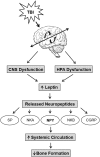
Traumatic brain injury (TBI) leads to long-term cognitive, behavioral, affective deficits, and increase neurodegenerative diseases. It is only in recent years that there is growing awareness that TBI even in its milder form poses long-term health consequences to not only the brain but to other organ systems. Also, the concept that hormonal signals and neural circuits that originate in the hypothalamus play key roles in regulating skeletal system is gaining recognition based on recent mouse genetic studies. Accordingly, many TBI patients have also presented with hormonal dysfunction, increased skeletal fragility, and increased risk of skeletal diseases. Research from animal models suggests that TBI may exacerbate the activation and inactivation of molecular pathways leading to changes in both osteogenesis and bone destruction. TBI has also been found to induce the formation of heterotopic ossification and increased callus formation at sites of muscle or fracture injury through increased vascularization and activation of systemic factors. Recent studies also suggest that the disruption of endocrine factors and neuropeptides caused by TBI may induce adverse skeletal effects. This review will discuss the long-term consequences of TBI on the skeletal system and TBI-induced signaling pathways that contribute to the formation of ectopic bone, altered fracture healing, and reduced bone mass.
Keywords: bone formation; bone resorption; fracture repair; growth hormone; heterotopic ossification; neuropeptides; osteoporosis.
Figures


Similar articles
-
The effect of traumatic brain injury on bone healing: an experimental study in a novel in vivo animal model.Injury. 2015 Apr;46(4):661-5. doi: 10.1016/j.injury.2015.01.044. Epub 2015 Jan 31. Injury. 2015. PMID: 25682315
-
Relationship between heterotopic ossification and traumatic brain injury: Why severe traumatic brain injury increases the risk of heterotopic ossification.J Orthop Translat. 2017 Nov 14;12:16-25. doi: 10.1016/j.jot.2017.10.002. eCollection 2018 Jan. J Orthop Translat. 2017. PMID: 29662775 Free PMC article. Review.
-
Post-traumatic hormonal disturbances: prolactin as a link between head injury and enhanced osteogenesis.J Endocrinol Invest. 1998 Feb;21(2):78-86. doi: 10.1007/BF03350319. J Endocrinol Invest. 1998. PMID: 9585380
-
The pathogenesis of heterotopic ossification after traumatic brain injury. A review of current literature.Acta Orthop Belg. 2020 Sep;86(3):369-377. Acta Orthop Belg. 2020. PMID: 33581019 Review.
-
Post-traumatic changes in insulin-like growth factor type 1 and growth hormone in patients with bone fractures and traumatic brain injury.Wien Klin Wochenschr. 2001 Feb 15;113(3-4):119-26. Wien Klin Wochenschr. 2001. PMID: 11253737
Cited by
-
Repeated mild traumatic brain injury impairs fracture healing in male mice.BMC Res Notes. 2022 Jan 29;15(1):25. doi: 10.1186/s13104-022-05906-7. BMC Res Notes. 2022. PMID: 35093144 Free PMC article.
-
Multi-omics analysis reveals GABAergic dysfunction after traumatic brainstem injury in rats.Front Neurosci. 2022 Nov 23;16:1003300. doi: 10.3389/fnins.2022.1003300. eCollection 2022. Front Neurosci. 2022. PMID: 36507346 Free PMC article.
-
Differential fracture response to traumatic brain injury suggests dominance of neuroinflammatory response in polytrauma.Sci Rep. 2019 Aug 21;9(1):12199. doi: 10.1038/s41598-019-48126-z. Sci Rep. 2019. PMID: 31434912 Free PMC article.
-
Bone Tissue and the Nervous System: What Do They Have in Common?Cells. 2022 Dec 22;12(1):51. doi: 10.3390/cells12010051. Cells. 2022. PMID: 36611845 Free PMC article. Review.
-
Determining the pharmacologic window of bisphosphonates that mitigates severe injury-induced osteoporosis and muscle calcification, while preserving fracture repair.Osteoporos Int. 2022 Apr;33(4):807-820. doi: 10.1007/s00198-021-06208-7. Epub 2021 Oct 31. Osteoporos Int. 2022. PMID: 34719727 Free PMC article.
References
-
- Centers for Disese Control and Prevention. Traumatic Brain Injury & Concussion. (2017). Available from: https://www.cdc.gov/traumaticbraininjury/index.html
Publication types
Grants and funding
LinkOut - more resources
Full Text Sources
Other Literature Sources

