. 2018 Apr;8(4):350-358.
doi: 10.3892/br.2018.1066. Epub 2018 Feb 16.

Characterization of human dental pulp cells grown in chemically defined serum-free medium

Affiliations

Characterization of human dental pulp cells grown in chemically defined serum-free medium

Sakiko Fujii et al. Biomed Rep. 2018 Apr.

Abstract

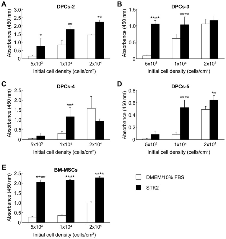
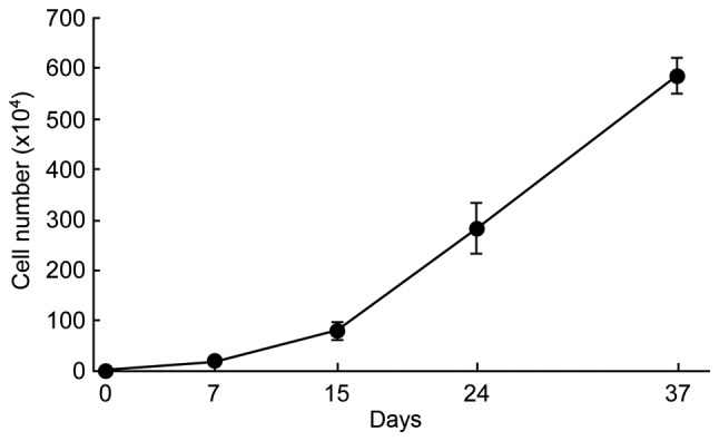
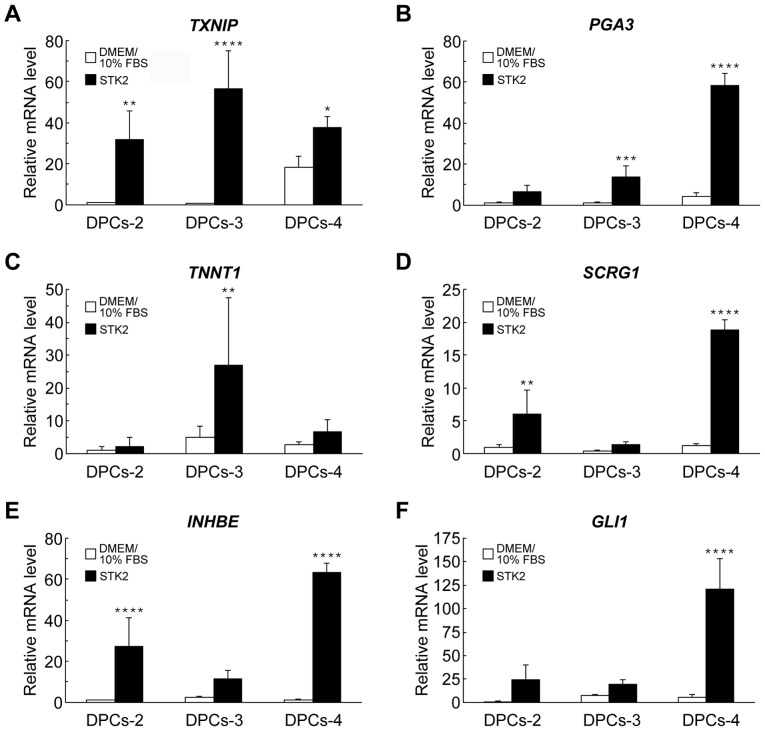
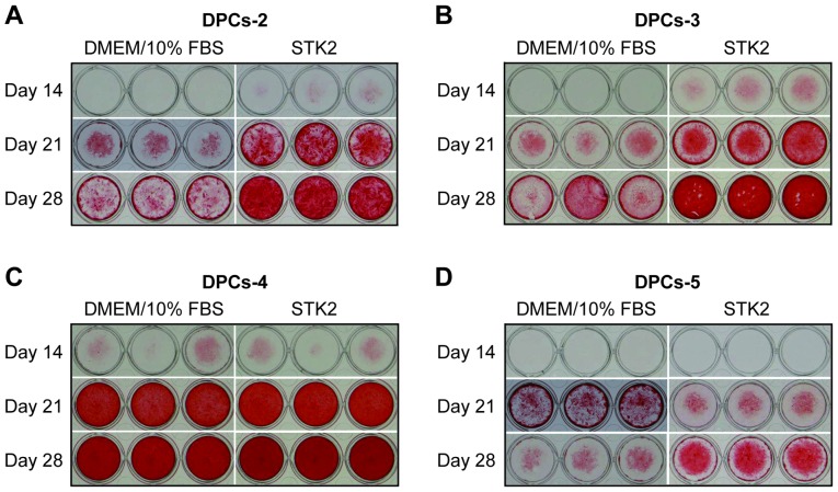
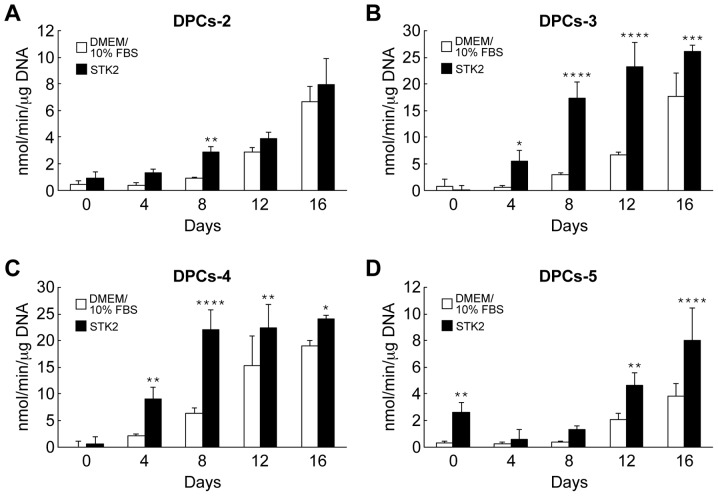
Dental pulp cells (DPCs) are promising candidates for use as transplantable cells in regenerative medicine. However, ex vivo expansion of these cells typically requires culture media containing fetal bovine serum, which may cause infection and immunological reaction following transplantation. In addition, the proliferation and differentiation of DPCs markedly depend upon serum batches. Therefore, the present study examined whether DPCs could be expanded under serum-free conditions. DPCs obtained from four donors were identified to proliferate actively in the serum-free medium, STK2, when compared with those cells in control medium (Dulbecco's modified Eagle's medium containing 10% serum). The high proliferative potential with STK2 was maintained through multiple successive culture passages. DNA microarray analyses demonstrated that the gene expression profile of DPCs grown in STK2 was similar to that of cells grown in the control medium; however, a number of genes related to cell proliferation, including placental growth factor and inhibin-βE, were upregulated in the STK2 cultures. Following induction of osteogenesis, DPCs grown in STK2 induced alkaline phosphatase activity and calcification at higher levels compared with the control medium cultures, indicating maintenance of differentiation potential in STK2. This serum-free culture system with DPCs may have applications in further experimental studies and as a clinical strategy in regenerative medicine.

Keywords: cell proliferation; dental pulp cells; gene expression; osteogenesis; regenerative medicine.


Figures

Figure 1.
Figure 1.
Effects of STK2 on the proliferation of four DPC lines. The DPC lines (A) DPCs-2, (B) DPCs-3, (C) DPCs-4 and (D) DPCs-5, and (E) BM-MSCs were seeded at the indicated initial densities and exposed to DMEM/10% FBS or STK2. Cell number was estimated on day 7 by Cell Counting Kit-8 assay. Values are the means ± standard deviation of at least three replicate cultures. *P<0.05, **P<0.01, ***P<0.001 and ****P<0.0001 vs. DMEM/10% FBS at the same cell density. DPCs, dental pulp cells; BM-MSCs, bone marrow-derived mesenchymal stem cells; DMEM/10% FBS, Dulbecco's modified Eagle's medium supplemented with 10% fetal bovine serum.
Figure 2.
Figure 2.
Growth curve of DPCs grown in STK2. DPCs-5 cells were seeded at low density (5×10³ cells/cm2) and exposed to STK2. When the cultures approached confluence, the cells were harvested with Accutase and a population was transferred into a new dish with STK2 to allow continued cell growth. The cumulative cell numbers are shown. Values are the means ± standard deviation of four replicate cultures. DPCs, dental pulp cells.
Figure 3.
Figure 3.
RT-qPCR analyses of gene expression in DPCs grown in DMEM/10% FBS or STK2. DPCs (DPCs-2, DPCs-3 and DPCs-4) were cultured in DMEM/10% FBS or STK2 for 7 days. The mRNA levels of (A) TXNIP, (B) PGA3, (C) TNNT1, (D) SCRG1, (E) INHBF and (F) GLI1 were quantified by RT-qPCR analysis. Values are the means ± standard deviation of four replicate cultures. *P<0.05, **P<0.01, ***P<0.001 and ****P<0.0001 vs. DMEM/10% FBS in the same cell line. DPCs, dental pulp cells; DMEM/10% FBS, Dulbecco's modified Eagle's medium supplemented with 10% fetal bovine serum; TXNIP, thioredoxin interacting protein; PGA3, pepsinogen 3; TNNT1, troponin T type 1; SCRG1, stimulator of chondrogenesis 1; INHBE, inhibin-βE; GLI1, GLI family zinc finger 1; RT-qPCR, reverse transcription-quantitative polymerase chain reaction.
Figure 4.
Figure 4.
Effects of pre-culture in STK2 on calcification in DPCs. The DPC lines (A) DPCs-2, (B) DPCs-3, (C) DPCs-4 and (D) DPCs-5 were seeded at low density (5×10³ cells/cm2) and pre-cultured for 7 days in DMEM/10% FBS or STK2. When cultures reached confluence, the cells were cultured in osteogenesis induction medium for up to 28 days. The calcified matrix was stained with Alizarin Red S on the indicated days following the exposure to osteogenesis induction medium. DPCs, dental pulp cells; DMEM/10% FBS, Dulbecco's modified Eagle's medium supplemented with 10% fetal bovine serum.
Figure 5.
Figure 5.
Effects of pre-culture in STK2 on ALP activity in DPCs. The dental pulp cell lines (A) DPCs-2, (B) DPCs-3, (C) DPCs-4 and (D) DPCs-5 were seeded at low density (5×10³ cells/cm2) and pre-cultured for 7 days in DMEM/10% FBS or STK2. When cultures reached confluence, the cells were cultured in osteogenesis induction medium for up to 16 days. ALP activity was determined on the indicated days following the exposure to osteogenesis induction medium. Values are the means ± standard deviation of four replicate cultures. *P<0.05, **P<0.01, ***P<0.001 and ****P<0.0001 vs. DMEM/10% FBS on the same day. ALP, alkaline phosphatase; DPCs, dental pulp cells; DMEM/10% FBS, Dulbecco's modified Eagle's medium supplemented with 10% fetal bovine serum.

References

    1. Gronthos S, Mankani M, Brahim J, Robey PG, Shi S. Postnatal human dental pulp stem cells (DPSCs) in vitro and in vivo; Proc Natl Acad Sci USA; 2000; pp. 13625–13630. - DOI - PMC - PubMed
    1. Stanislawski L, Carreau JP, Pouchelet M, Chen ZH, Goldberg M. In vitro culture of human dental pulp cells: Some aspects of cells emerging early from the explant. Clin Oral Investig. 1997;1:131–140. doi: 10.1007/s007840050024. - DOI - PubMed
    1. Couble ML, Farges JC, Bleicher F, Perrat-Mabillon B, Boudeulle M, Magloire H. Odontoblast differentiation of human dental pulp cells in explant cultures. Calcif Tissue Int. 2000;66:129–138. doi: 10.1007/PL00005833. - DOI - PubMed
    1. He H, Yu J, Liu Y, Lu S, Liu H, Shi J, Jin Y. Effects of FGF2 and TGFbeta1 on the differentiation of human dental pulp stem cells in vitro. Cell Biol Int. 2008;32:827–834. doi: 10.1016/j.cellbi.2008.03.013. - DOI - PubMed
    1. Iohara K, Zheng L, Ito M, Tomokiyo A, Matsushita K, Nakashima M. Side population cells isolated from porcine dental pulp tissue with self-renewal and multipotency for dentinogenesis, chondrogenesis, adipogenesis, and neurogenesis. Stem Cells. 2006;24:2493–2503. doi: 10.1634/stemcells.2006-0161. - DOI - PubMed