

Case Reports
doi: 10.1016/j.eucr.2018.03.005.
eCollection 2018 May.
Vesical fungus balls (fungal bezoars) by Candida albicans mimicking urothelial carcinoma in a patient with diabetic neurogenic bladder
Affiliations
- PMID: 29556476
- PMCID: PMC5854892
- DOI: 10.1016/j.eucr.2018.03.005
Item in Clipboard
Case Reports
Vesical fungus balls (fungal bezoars) by Candida albicans mimicking urothelial carcinoma in a patient with diabetic neurogenic bladder
Urol Case Rep.
.
No abstract available
Keywords: Candida albicans; Diabetes complications; Magnetic resonance imaging; Urinary bladder.
Figures
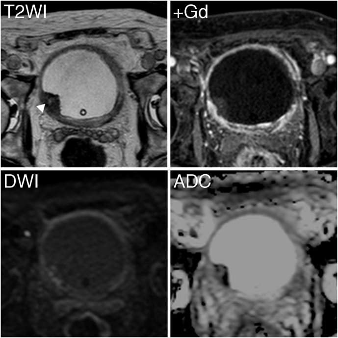
Magnetic resonance imaging findings of the pre-therapeutic urinary bladder lesion are shown. The lesion showed hypointensity for the most part despite a small portion with hyperintensity indicating necrotic portion (arrowed) on T2WI, T2-weighted imaging; enhancement on +Gd, Gadolinium-enhanced imaging; restriction on DWI, diffusion-weighted imaging; a low value on ADC, apparent diffusion coefficient mapping.
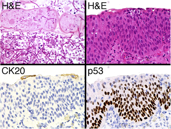
Microscopic photographs of the resected tissues are shown. Hyphae of Candida albicans were observed among degenerated epithelial cells (upper left panel; H&E, hematoxylin and eosin stain). Nuclei of the background urinary bladder epithelial cells were enlarged and irregular; however, structural atypia did not reach the degree seen in cancer cells (upper right panel). Moreover, CK20-positivity was limited in the cytoplasm of umbrella cells (lower left panel) and p53-positivity was weak in the nuclei of basal cells (lower right panel).
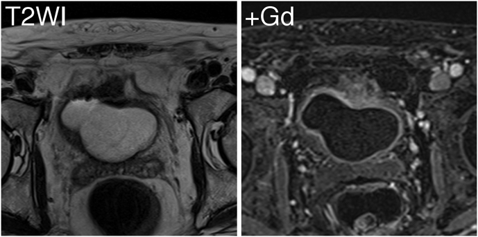
Magnetic resonance imaging findings of the post-therapeutic urinary bladder lesion are shown. There was no mass lesion observed in the bladder on T2WI, T2-weighted imaging; nor was there significant enhancement of the thickening wall of the bladder on +Gd, Gadolinium-enhanced imaging.
Similar articles
-
Acute renal failure as a result of bilateral ureteral obstruction by Candida albicans fungus balls.Int J Urol. 2006 Aug;13(8):1121-2. doi: 10.1111/j.1442-2042.2006.01509.x. Int J Urol. 2006. PMID: 16903942
-
[Bilateral candida bezoar of the upper urinary tract].Actas Urol Esp. 1998 Apr;22(4):374-6. Actas Urol Esp. 1998. PMID: 9658653 Spanish.
-
Case report of a ureteral obstruction by Candida albicans fungus balls detected by magnetic resonance imaging in kidney transplant recipient.Exp Clin Transplant. 2014 Dec;12(6):559-61. doi: 10.6002/ect.2013.0235. Epub 2014 Jun 25. Exp Clin Transplant. 2014. PMID: 25020145
-
[Candida urinary tract infection with special reference to ascending pyelonephritis].Hinyokika Kiyo. 1991 Sep;37(9):969-74. Hinyokika Kiyo. 1991. PMID: 1785422 Review. Japanese.
-
Role of diffusion-weighted magnetic resonance imaging as an imaging biomarker of urothelial carcinoma.Int J Urol. 2014 Dec;21(12):1190-200. doi: 10.1111/iju.12587. Epub 2014 Jul 30. Int J Urol. 2014. PMID: 25074594 Review.
Cited by
-
Vesical fungal bezoars on post-TURBT scar tissue causing obstruction and mimicking urothelial carcinoma.BMJ Case Rep. 2024 Jan 12;17(1):e257132. doi: 10.1136/bcr-2023-257132. BMJ Case Rep. 2024. PMID: 38216163
-
Fungal Bezoars Mimicking an Enterovesica Fistula: A Unique Case Report.Curr Urol. 2019 Oct;13(2):107-109. doi: 10.1159/000499284. Epub 2019 Oct 1. Curr Urol. 2019. PMID: 31768178 Free PMC article.
-
Imaging spectrum of common and rare infections affecting the lower genitourinary tract.Abdom Radiol (NY). 2021 Jun;46(6):2665-2682. doi: 10.1007/s00261-020-02889-6. Epub 2021 Jan 3. Abdom Radiol (NY). 2021. PMID: 33388810 Review.
-
Imaging of non-traumatic urinary bladder emergencies.Emerg Radiol. 2019 Dec;26(6):675-682. doi: 10.1007/s10140-019-01703-1. Epub 2019 Jul 6. Emerg Radiol. 2019. PMID: 31280426 Review.
-
Male with urinary urgency, frequency, and dysuria.J Am Coll Emerg Physicians Open. 2022 Mar 14;3(2):e12696. doi: 10.1002/emp2.12696. eCollection 2022 Apr. J Am Coll Emerg Physicians Open. 2022. PMID: 35316969 Free PMC article. No abstract available.
References
-
- Chisholm E.R., Hutch J.A. Fungus ball (Candida albicans) formation in the bladder. J Urol. 1961;86:559–562. - PubMed
-
- Irby P.B., Stoller M.L., McAninch J.W. Fungal bezoars of the upper urinary tract. J Urol. 1990;143(3):447–451. - PubMed
-
- Mallofré C., Castillo M., Morente V., Solé M. Immunohistochemical expression of CK20, p53, and Ki-67 as objective markers of urothelial dysplasia. Mod Pathol. 2003;16(3):187–191. - PubMed
-
- Kauffman C.A., Vazquez J.A., Sobel J.D. Prospective multicenter surveillance study of funguria in hospitalized patients. The national institute for allergy and infectious diseases (NIAID) mycoses study group. Clin Infect Dis. 2000;30(1):14–18. - PubMed
-
- Fisher J.F. Candida urinary tract infections–epidemiology, pathogenesis, diagnosis, and treatment: executive summary. Clin Infect Dis. 2011;52(Suppl 6):S429–S432. - PubMed
Publication types
LinkOut - more resources
Full Text Sources
Other Literature Sources
