. 2018 Feb 16:18:17-24.
doi: 10.1016/j.ijcha.2017.12.003. eCollection 2018 Mar.

Mobilization of progenitor cells and assessment of vessel healing after second generation drug-eluting stenting by optical coherence tomography

Affiliations

Mobilization of progenitor cells and assessment of vessel healing after second generation drug-eluting stenting by optical coherence tomography

Masashi Sakuma et al. Int J Cardiol Heart Vasc. .

Erratum in

Abstract

Background: Bone marrow-derived progenitor cells likely contribute to both endothelial- and smooth muscle cell-dependent healing responses in stent-injured vessel sites. This study aimed to assess mobilization of progenitor cells and vessel healing after zotarolimus-eluting (ZES) and everolimus-eluting (EES) stents.

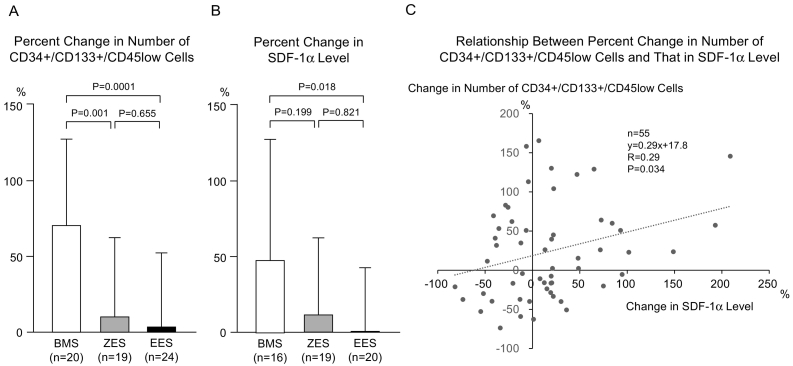
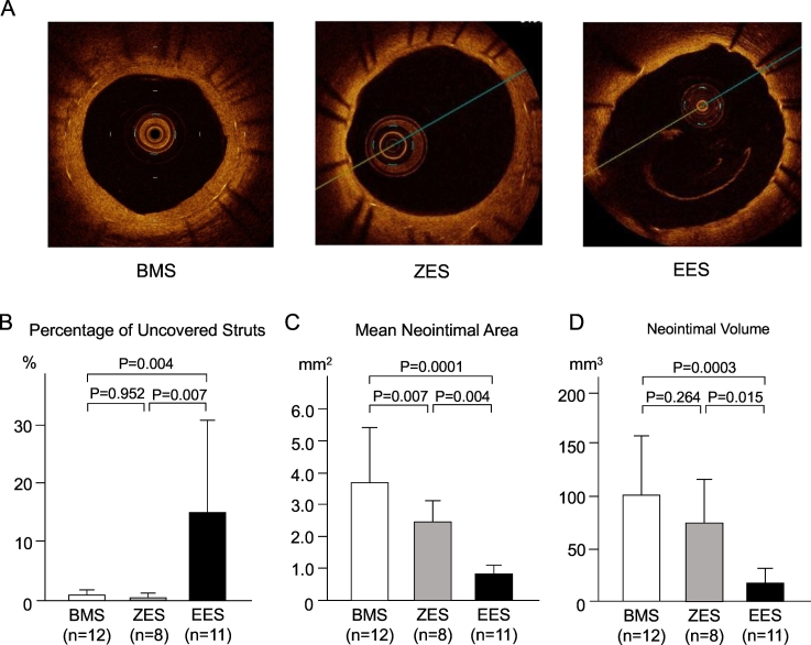
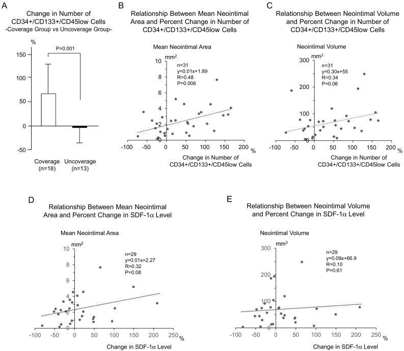
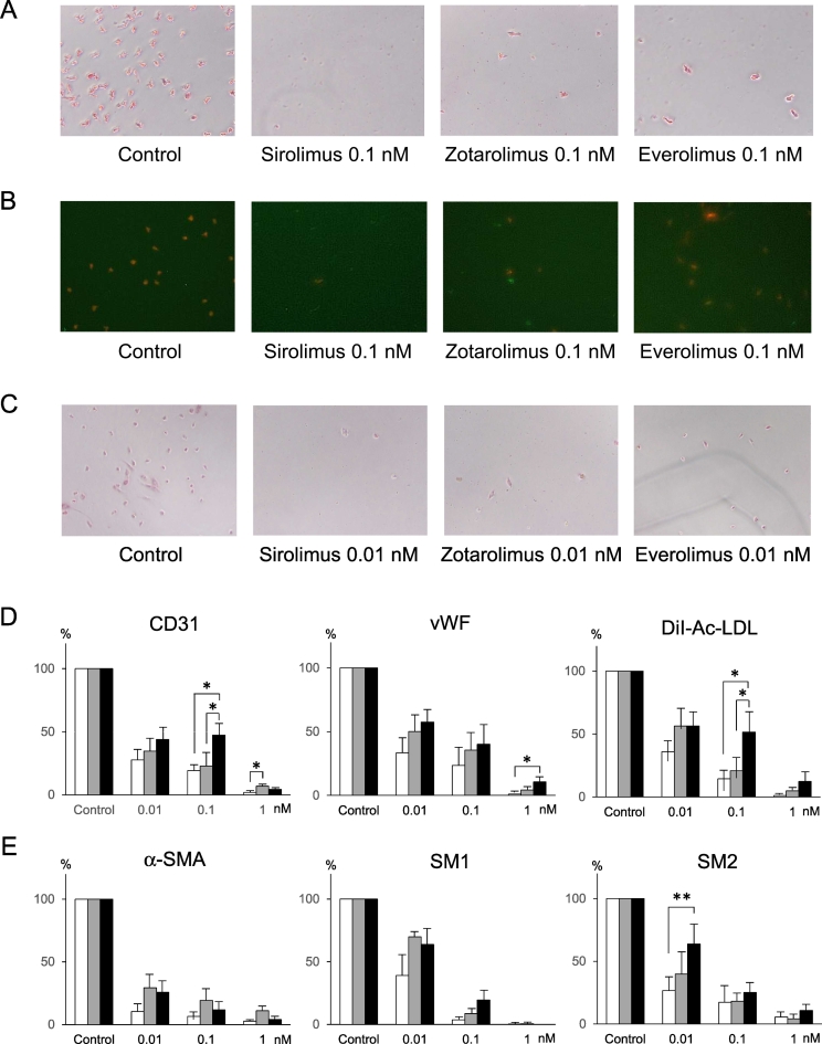
Methods and results: In 63 patients undergoing coronary stent implantation, we measured circulating CD34 + CD133 + CD45low cells and serum levels of biomarkers relevant to stem cell mobilization. In 31 patients of them, we assessed vessel healing within the stented segment using optical coherence tomography (OCT) imaging. The CD34 + CD133 + CD45low cells increased 68 ± 59% 7 days after bare metal stent (BMS), 10 ± 53% after ZES (P < 0.01 vs BMS), 3 ± 49% after EES (P < 0.001 vs BMS), compared with baseline. Percent change in CD34 + CD133 + CD45low cells was positively correlated with that in stromal cell-derived factor (SDF)-1α (R = 0.29, P = 0.034). Percentage of uncovered struts was higher in the EES group (14.4 ± 17.3%), compared with the BMS (0.7 ± 1.3, P < 0.01) and ZES (0.4 ± 0.5, P < 0.01) groups. The change in CD34 + CD133 + CD45low cells showed positive correlation with OCT-quantified mean neointimal area (R = 0.48, P < 0.01). Finally, circulating mononuclear cells obtained from 5 healthy volunteers were isolated to determine the effect of sirolimus, zotarolimus and everolimus on vascular cell differentiation. The differentiation of mononuclear cells into endothelial-like cells was dose-dependently suppressed by sirolimus, zotarolimus, and everolimus.

Conclusions: Mobilization of progenitor cells was suppressed, and differentiation of mononuclear cells into endothelial-like cells was inhibited, in association with increased number of uncovered stent struts, even after second generation drug-eluting stenting. These data suggest that new approaches are necessary to enhance stent healing.

Keywords: Circulating progenitor cell; Drug-eluting stent; Optical coherence tomography; Re-endothelialization; Vascular injury.


Figures

Fig. 1
Fig. 1
Circulating CD34 + CD133 + CD45low cells and SDF-1α level. (A) Percent change in the number of CD34 + CD133 + CD45low cells at the day 7 from the baseline value was less in the ZES group and was still less in the EES group, compared with the BMS group. (B) Percent change in the SDF-1α level was less in the EES group, compared with the BMS group. The level in the ZES group did not show significant difference from that in BMS group as well as that in EES group. (C) In overall patients, the percent change in number of CD34 + CD133 + CD45low cells was positively correlated with that in SDF-1α level. BMS indicates bare metal stent; ZES, zotarolimus-eluting stent; EES, everolimus-eluting stent; SDF, stromal cell-derived factor.
Fig. 2
Fig. 2
OCT-based neointima formation and circulating CD34 + CD133 + CD45low cells. (A) Typical OCT findings indicate that neointima formation and neointimal coverage over the stent struts are adequate in a BMS lesion, a little inferior in a ZES lesion and poor in an EES lesion. (B) Percentage of uncovered struts was higher in the EES group, compared with the BMS group and ZES group. (C) Mean neointimal area was less in EES group, compared with the BMS group and ZES group. The value in the EES group was still less than that in the ZES group. (D) Neointimal volume was less in the EES group, compared with the BMS group and ZES group.
Fig. 3
Fig. 3
Neointima formation vs circulating CD34 + CD133 + CD45low cells and SDF-1α level. (A) If the patients were divided into 2 groups, i.e., coverage group (percent uncovered struts < 1.0%) or uncoverage group (1.0%≤), independently of stent type, percent changes in the number of CD34 + CD133 + CD45low cells were greater in the coverage group, compared with the uncoverage group. (B) Percent change in the number of CD34 + CD133 + CD45low cells showed significant positive correlation with the mean neointimal area. (C) Percent change in the number of CD34 + CD133 + CD45low cells showed a trend of positive correlation with the neointimal volume. (D) Percent change in SDF-1α level showed a trend of positive correlation with the mean neointimal area. (E) Percent change in SDF-1α level was not correlated with the neointimal volume.
Fig. 4
Fig. 4
Effect of sirolimus, zotarolimus and everolimus on differentiation of progenitor cells into endothelilal- and smooth muscle-like cells. (A) Sirolimus, zotarolimus and everolimus (0.1 nM each) reduced differentiation into CD31-positive endothelial-like cells, compared with control. The reduction was a little weaker in zotarolimus and everolimus, compared with sirolimus. (B) Compared with control, sirolimus, zotarolimus and everolimus (0.1 nM each) reduced differentiation into DiI-Ac-LDL/FITC-BS lectin-double positive cells, shown as orange-colored cells, in which red fluorescence (DiI) and green fluorescence (FITC) were merged. The reduction was a little weaker in zotarolimus and everolimus, compared with sirolimus. (C) Sirolimus, zotarolimus and everolimus (0.01 nM each) reduced differentiation into α-SMA-positive endothelial-like cells, compared with control. (D) Sirolimus, zotarolimus and everolimus significantly inhibited differentiation into endothelial-like cells, concentration-dependently. The inhibition seemed a little less in the everolimus, compared with sirolimus and zotarolimus. (E) Sirolimus, zotarolimus and everolimus significantly inhibited differentiation into smooth muscle-like cells, concentration-dependently. The inhibition seemed a little less in the everolimus, compared with sirolimus. vWF indicates von Willebrand factor; α-SMA, α-smooth muscle actin. formula image sirolimus formula imagezotarolimus formula imageeverolimus *P < 0.05, **P < 0.01.

References

    1. Kereiakes D.J. The TWENTE trial in perspective: stents and stent trials in evolution. JAMA Cardiol. 2017;2:235–237. - PubMed
    1. Inoue T., Node K. Molecular basis of restenosis and novel issues of drug-eluting stents. Circ. J. 2009;73:615–621. - PubMed
    1. Inoue T., Croce K., Morooka T., Sakuma M., Node K., Simon D.I. Vascular inflammation and repair: implication for reendothelialization, restenosis, and stent thrombosis. J. Am. Coll. Cardiol. Intv. 2011;4:1057–1066. - PMC - PubMed
    1. Costa M.A., Simon D.I. Molecular basis of restenosis and drug-eluting stents. Circulation. 2005;111:2257–2273. - PubMed
    1. Asahara T., Murohara T., Sullivan A., Silver M., van der Zee R., Li T., Witzenbichler B., Schatteman G., Isner J.M. Isolation of putative progenitor endothelial cells for angiogenesis. Science. 1997;275:964–966. - PubMed

LinkOut - more resources