Review
. 2018 Mar;12(1):13-21.
doi: 10.1007/s12105-018-0896-4. Epub 2018 Mar 20.

Proceedings of the NASHNP Companion Meeting, March 18th, 2018, Vancouver, BC, Canada: Salivary Neuroendocrine Carcinoma-An Overview of a Rare Disease with an Emphasis on Determining Tumor Origin

Affiliations
Review

Proceedings of the NASHNP Companion Meeting, March 18th, 2018, Vancouver, BC, Canada: Salivary Neuroendocrine Carcinoma-An Overview of a Rare Disease with an Emphasis on Determining Tumor Origin

Rebecca D Chernock et al. Head Neck Pathol. 2018 Mar.

Abstract

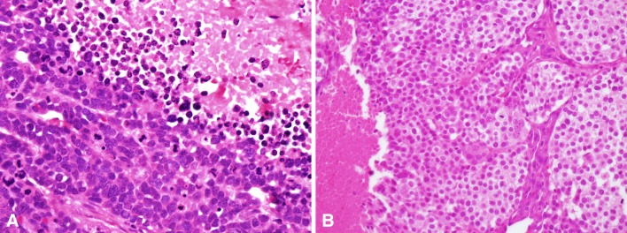
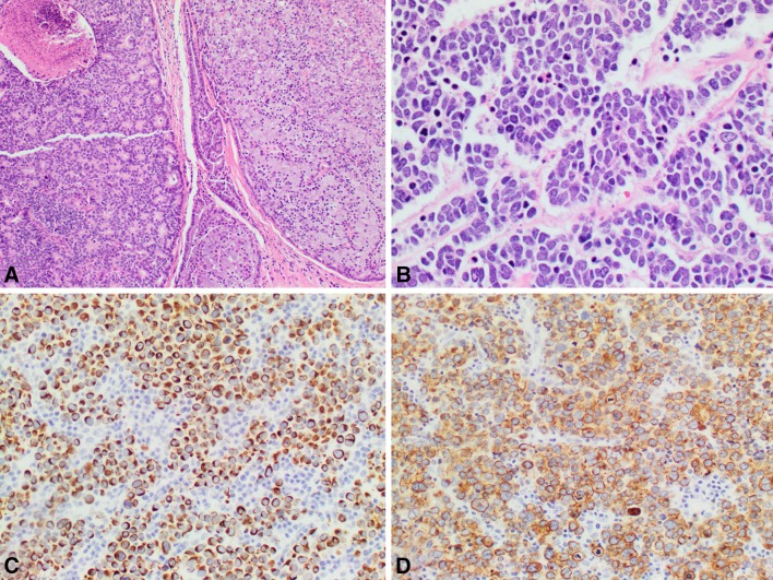
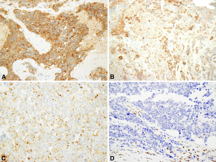
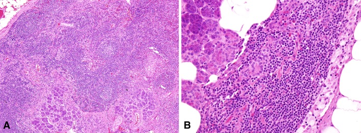
Salivary neuroendocrine carcinomas are rare and the overwhelming majority is high-grade. The parotid gland is the most commonly involved site followed by the submandibular gland. Most arise de novo but rare examples occurring as a high-grade transformation of another type of salivary gland neoplasm exist. There is significant morphologic and immunophenotypic overlap with neuroendocrine carcinomas of other sites, especially the skin. Like cutaneous neuroendocrine (or Merkel cell) carcinomas, approximately three-fourths are cytokeratin 20 positive. Cytokeratin 20 positive salivary neuroendocrine carcinomas are often referred to as being of the 'Merkel cell type' since most other non-cutaneous neuroendocrine carcinomas are cytokeratin 20 negative. Salivary neuroendocrine carcinomas may be challenging to separate from Merkel cell carcinomas of the head and neck on pathologic grounds because the latter often metastasize to the parotid gland. Clinical history is often relied upon to separate primary salivary tumors from cutaneous metastases but may not be helpful in all cases. Here we review the clinical, pathologic and molecular features of salivary neuroendocrine carcinomas focusing on high-grade major salivary gland tumors. The difficulty in separating salivary tumors from metastatic Merkel cell carcinoma will be highlighted.

Keywords: Large cell neuroendocrine carcinoma; Merkel cell carcinoma; Merkel cell polyomavirus; Neuroendocrine carcinoma; Salivary; Small cell carcinoma.


Conflict of interest statement

The authors have no sources of funding or conflicts of interest to disclose.

Figures

Fig. 1
Fig. 1
Examples of small and large cell salivary neuroendocrine carcinomas. Small cell carcinoma (a, ×600 magnification) is composed of sheets, nest and trabeculae of cells that have high nuclear to cytoplasmic ratios, finely granular chromatin, scant cytoplasm and often nuclear molding. Necrosis is present (top) and mitotic activity is brisk. Large cell neuroendocrine carcinoma (b, ×400 magnification) contains larger cells with more abundant cytoplasm and in some case prominent nucleoli
Fig. 2
Fig. 2
Small cell carcinoma arising from acinic cell carcinoma as a form of high grade transformation. A high grade component is present (left) with necrosis adjacent to the low grade acinic cell carcinoma (right, a, ×100 magnification). Typical features of small cell carcinoma including hyperchromatic nuclei, high nuclear to cytoplasmic ratios, nuclear molding and apoptotic debris are seen in the high grade component (b, ×400 magnification). The tumor cells are positive for cytokeratin 20 (c, ×200 magnification) and chromogranin (d, ×200 magnification). Images courtesy of Dr. Lester D. Thompson
Fig. 3
Fig. 3
Immunophenotype of salivary neuroendocrine carcinomas. One or more neuroendocrine markers (a, synaptophysin; b, chromogranin, ×200 magnification) are typically positive. Cytokeratin 20 positivity (c, ×200 magnification) is frequently observed, akin to cutaneous Merkel cell carcinomas, and pRB expression is often lost (d, ×400 magnification)
Fig. 4
Fig. 4
Salivary inclusions in parotid lymph nodes. Parotid lymph nodes frequently contain intranodal salivary tissue (a, ×100 magnification; b, ×400 magnification) that may be intraparenchymal or subcapsular

References

    1. Nagao T, Gaffey TA, Olsen KD, Serizawa H, Lewis JE. Small cell carcinoma of the major salivary glands: clinicopathologic study with emphasis on cytokeratin 20 immunoreactivity and clinical outcome. Am J Surg Pathol. 2004;28:762–770. doi: 10.1097/01.pas.0000126776.65815.48. - DOI - PubMed
    1. Gnepp DR, Wick MR. Small cell carcinoma of the major salivary glands. An immunohistochemical study. Cancer. 1990;66:185–192. doi: 10.1002/1097-0142(19900701)66:1<185::AID-CNCR2820660133>3.0.CO;2-4. - DOI - PubMed
    1. Mair S, Phillips JI, Cohen R. Small cell undifferentiated carcinoma of the parotid gland. Cytologic, histologic, immunohistochemical and ultrastructural features of a neuroendocrine variant. Acta Cytol. 1989;33:164–168. - PubMed
    1. de Vicente Rodríguez JC, Fresno Forcelledo MF, Junquera Gutiérrez LM, Hernández Vallejo G, López Arranz JS. Small cell undifferentiated carcinoma of the submandibular gland with neuroendocrine features. Ann Otol Rhinol Laryngol. 2004;113:55–59. doi: 10.1177/000348940411300113. - DOI - PubMed
    1. Mulder DC, Rosenberg AJWP, Storm-Bogaard PW, Koole R. Spontaneous regression of advanced merkel-cell-like small cell carcinoma of the parotid gland. Br J Oral Maxillofac Surg. 2010;48:199–200. doi: 10.1016/j.bjoms.2009.06.008. - DOI - PubMed

LinkOut - more resources