Case Reports
. 2018 Mar;48(3):236-239.
doi: 10.4070/kcj.2017.0133.

Isolated Cardiac Sarcoidosis Presenting with Stroke

Affiliations
Case Reports

Isolated Cardiac Sarcoidosis Presenting with Stroke

Masanari Kuwabara et al. Korean Circ J. 2018 Mar.
No abstract available


Conflict of interest statement

The authors have no financial conflicts of interest.

Figures

Figure 1
Figure 1
Brain magnetic resonance image. This image shows multiple cerebrovascular infarctions especially in the cerebellum.
Figure 2
Figure 2
Echocardiography shows LV thrombus. The thorombus is 2.64×1.74 cm in the LV apical lesion. LV = left ventricular.
Figure 3
Figure 3
Coronary CTA. This image shows normal coronary arteries. CTA = computed tomography angiography.
Figure 4
Figure 4
Gallium-67 scintigraphy. This image shows no specific lesion in the body.
Figure 5
Figure 5
Cardiac MRI image. (A) T2WI and (B) LGE image. There are multiple focal LGEs in left ventricle (B) where T2WI also shows pale high intensity with unclear margin. It suggests active inflammation. LGE = late gadolinium enhancement; MRI = magnetic resonance imaging; T2WI = T2 star weighted image.
Figure 6
Figure 6
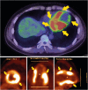
Fasting 18F-fluorodeoxyglucose PET. The image shows multiple increased cardiac uptake lesions, especially at the anterior, basal lateral, and inferior LV lesions. The contrast is clear, and the lesions are very similar to LGE lesions. However, apical lesions show low uptake, which suggests scar lesions. LGE = late gadolinium enhancement; LV = left ventricular; PET = positron emission tomography.

Similar articles

Cited by

References

    1. Sadek MM, Yung D, Birnie DH, Beanlands RS, Nery PB. Corticosteroid therapy for cardiac sarcoidosis: a systematic review. Can J Cardiol. 2013;29:1034–1041. - PubMed
    1. Brown MM, Thompson AJ, Wedzicha JA, Swash M. Sarcoidosis presenting with stroke. Stroke. 1989;20:400–405. - PubMed
    1. White J, Sutton T, Kerr A. Isolated primary cardiac sarcoidosis: MRI diagnosis and monitoring of treatment response with cardiac enzymes. Circ Heart Fail. 2010;3:e28–9. - PubMed
    1. Isobe M, Tezuka D. Isolated cardiac sarcoidosis: clinical characteristics, diagnosis and treatment. Int J Cardiol. 2015;182:132–140. - PubMed
    1. Takaya Y, Kusano KF, Nakamura K, Ito H. Comparison of outcomes in patients with probable versus definite cardiac sarcoidosis. Am J Cardiol. 2015;115:1293–1297. - PubMed

Publication types

LinkOut - more resources