Review
. 2018 Aug;24(4):381-399.
doi: 10.1177/1073858418760481. Epub 2018 Mar 20.

Neural Mechanisms of Inflammation-Induced Fever

Affiliations
Review

Neural Mechanisms of Inflammation-Induced Fever

Anders Blomqvist et al. Neuroscientist. 2018 Aug.

Abstract

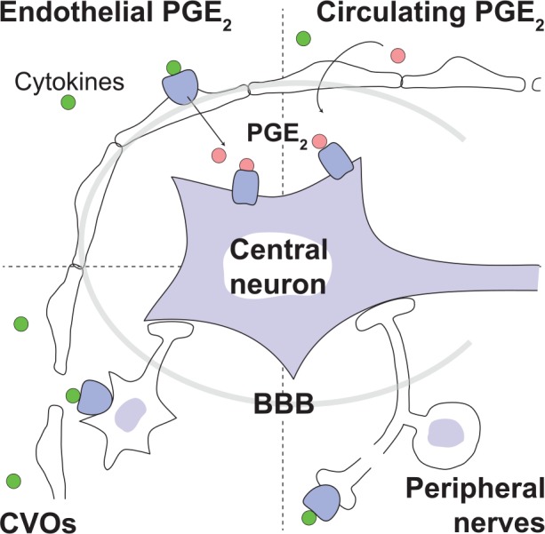
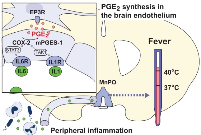
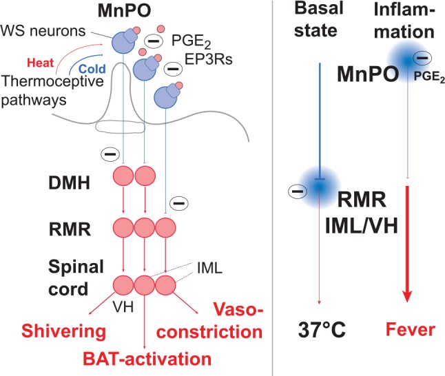
Fever is a common symptom of infectious and inflammatory disease. It is well-established that prostaglandin E2 is the final mediator of fever, which by binding to its EP3 receptor subtype in the preoptic hypothalamus initiates thermogenesis. Here, we review the different hypotheses on how the presence of peripherally released pyrogenic substances can be signaled to the brain to elicit fever. We conclude that there is unequivocal evidence for a humoral signaling pathway by which proinflammatory cytokines, through their binding to receptors on brain endothelial cells, evoke fever by eliciting prostaglandin E2 synthesis in these cells. The evidence for a role for other signaling routes for fever, such as signaling via circumventricular organs and peripheral nerves, as well as transfer into the brain of peripherally synthesized prostaglandin E2 are yet far from conclusive. We also review the efferent limb of the pyrogenic pathways. We conclude that it is well established that prostaglandin E2 binding in the preoptic hypothalamus produces fever by disinhibition of presympathetic neurons in the brain stem, but there is yet little understanding of the mechanisms by which factors such as nutritional status and ambient temperature shape the response to the peripheral immune challenge.

Keywords: EP3 receptors; brain endothelial cells; cytokines; fever; median preoptic nucleus; prostaglandin E2.


Conflict of interest statement

Declaration of Conflicting Interests: The author(s) declared no potential conflicts of interest with respect to the research, authorship, and/or publication of this article.

Figures

Figure 1.
Figure 1.
Different suggested routes by which peripherally released inflammatory signals can bypass the blood-brain barrier (BBB) and activate the central nervous system: Peripherally released proinflammatory cytokines (green circles) (i) bind to receptors on cells of brain blood vessels to induce synthesis of prostaglandin E2 (PGE2; pink circles), which then is transported into the brain parenchyma (upper left); (ii) activate neurons of circumventricular organs (CVOs), which contain fenestrated capillaries (lower left); or (iii) activate peripheral nerves (lower right). (iv) Peripheral inflammation may also release circulating PGE2 that enters the brain (upper right).
Figure 2.
Figure 2.
Confocal micrographs of blood vessel in the mouse brain, stained with antibodies against the prostaglandin E2 synthesizing enzymes cyclooxygenase-2 (Cox-2) and microsomal prostaglandin E synthase-1 (mPGES-1), and CD206, a macrophage marker expressed by perivascular cells. Upper left panel shows triple labeling for these proteins, and the other panels single labeling for each protein. Note that most cells that express Cox-2 also express mPGES-1 and vice versa. Note also that none of the Cox-2/mPGES-1 expressing cells stain for CD206, implying that this population does not include perivascular cells. Scale bar = 20 µm.
Figure 3.
Figure 3.
Temperature responses to intraperitoneal injection of bacterial wall lipopolysaccharide (LPS) in wild type (WT) and mPGES-1 knockout (KO) mice that were subjected to whole body irradiation and then transplanted with either WT (+/+) or KO (−/−) bone marrow. Note that WT mice (non-BM +/+) transplanted with WT or KO bone marrow display a prominent febrile response (two top fever curves), whereas KO mice (non-BM −/−) transplanted with WT or KO bone marrow are afebrile (lower fever curves). The initial temperature peak (shadowed) in all groups is handling stress-induced hyperthermia. Replacement of native hematopoietically derived cells was in these experiments about 90% among white blood cells and brain macrophages (perivascular cells), and around 70% among liver (Kupffer cells) and lung macrophages. For the NaCl treated group mean is shown, whereas for the other traces mean and SEM (standard error of the mean) are shown. Adapted from Engström and others (2012).
Figure 4.
Figure 4.
Blunted febrile response to intraperitoneally injected bacterial wall lipopolysaccharide (LPS) in mice with deletion selectively in brain endothelial cells of the prostaglandin E2 synthesizing enzymes cyclooxygenase-2 (Cox2ΔbEnd) and microsomal prostaglandin E synthase-1 (mPGES1ΔbEnd). WT, wild type mice. For the NaCl-treated groups mean is shown, whereas for the LPS treated groups mean and SEM (standard error of the mean) are shown. Adapted from Wilhelms and others (2014).
Figure 5.
Figure 5.
Febrile response to bacterial wall lipopolysaccharide (LPS) in mice with deletion of cytokine receptors. (A) Mice with global deletion of the interleukin-1 type 1 receptor (IL1R-KO) show attenuated fever, however note the late appearing fever in these mice. (B) Attenuated fever, seen after about 5 h, in mice with deletion of the IL-1R1 selectively in brain endothelial cells (IL1RΔbEnd). (C) Attenuated fever in mice with deletion of the interleukin-6 receptor alpha selectively in brain endothelial cells (IL6RΔbEnd) (left). This response was associated with attenuated induction of cyclooxygenase-2 (Cox-2) in the hypothalamus (right). ** indicates P < 0.01. Adapted from Matsuwaki and others (2017) and Eskilsson and others (2014).
Figure 6.
Figure 6.
Transduction mechanisms in the blood-brain barrier elicited by peripherally released inflammatory mediators. The cytokines IL-1β and IL-6 (green circles) bind to receptors (IL1R, IL6R) on brain endothelial cells in the preoptic hypothalamus resulting in transcription of cyclooxygenase-2 (COX-2) and microsomal prostaglandin E synthase-1 (mPGES-1) via TAK1 and STAT3, respectively. The subsequent binding of neosynthesized PGE2 (pink circles) to PGE2 EP3 receptor (EP3R) expressing cells in the median preoptic nucleus (MnPO) of the hypothalamus elicits fever.
Figure 7.
Figure 7.
Fever response to intravenously injected lipopolysaccharide (LPS) in whole-body irradiated wild type (WT) and mPGES-1 knockout (KO) mice transplanted with WT (+/+) and KO (−/−) bone marrow. WT mice (non-BM +/+) show a first phase of fever, irrespective of whether they were transplanted with WT or KO bone marrow (upper two traces; cf. the temperature curve for mice injected with saline). In contrast, KO mice (non-BM −/−) transplanted with WT bone marrow instead show a hypothermic response, similar to KO mice transplanted with KO bone marrow (lower two traces). For all traces mean is shown. Dashed vertical line indicates time of injection. For further details of these experiments, see Figure 3. Adapted from Engström and others (2012).
Figure 8.
Figure 8.
Circuitry of pyrogenic pathways from the preoptic hypothalamus. EP3 receptor expressing neurons in the median preoptic nucleus (MnPO) exert tonic inhibition on presympathetic neurons in the rostral medullary raphe nucleus (RMR) as well as on neurons in the dorsomedial hypothalamus (DMH), hence silencing, in the resting state, sympathetic, thermogenic output from the intermediolateral cell column (IML) as well as excitatory output to motorneurons in the ventral horn (VH) of the spinal cord responsible for shivering thermogenesis. On immune-induced PGE2 release (pink circles) and binding to the EP3 receptors (EP3Rs), the MnPO neurons are silenced, resulting in disinhibition of neurons in DMH and RMR and activation of the thermogenic circuitry. In addition to responding to PGE2, EP3 expressing neurons are warm-sensitive (WS), and hence activated by heat, inhibiting thermogenesis, and inhibited by cold, promoting thermogenesis. Note that the direct projection from MnPO to RMR controls vasoconstriction, whereas the indirect pathway over the DMH controls shivering and non-shivering (brown adipose tissue [BAT] activation) thermogenesis.

Similar articles

Cited by

References

    1. Abbott SBG, Machado NLS, Geerling JC, Saper CB. 2016. Reciprocal control of drinking behavior by median preoptic neurons in mice. J Neurosci 36(31):8228–37. - PMC - PubMed
    1. Alheim K, Chai Z, Fantuzzi G, Hasanvan H, Malinowsky D, Di Santo E, and others. 1997. Hyperresponsive febrile reactions to interleukin (IL) 1α and IL-1β, and altered brain cytokine mRNA and serum cytokine levels, in IL-1β-deficient mice. Proc Natl Acad Sci U S A 94(6):2681–6. - PMC - PubMed
    1. Almeida MC, Steiner AA, Branco LG, Romanovsky AA. 2006. Neural substrate of cold-seeking behavior in endotoxin shock. PLoS One 1:e1. - PMC - PubMed
    1. Atkins E. 1982. Fever: Its history, cause, and function. Yale J Biol Med 55(3–4):283–9. - PMC - PubMed
    1. Binshtok AM, Wang H, Zimmermann K, Amaya F, Vardeh D, Shi L, and others. 2008. Nociceptors are interleukin-1β sensors. J Neurosci 28(52):14062–73. - PMC - PubMed

Publication types