

Hyperhomocysteinemia induces injury in olfactory bulb neurons by downregulating Hes1 and Hes5 expression
- PMID: 29557377
- PMCID: PMC5879899
- DOI: 10.4103/1673-5374.220779
Hyperhomocysteinemia induces injury in olfactory bulb neurons by downregulating Hes1 and Hes5 expression
Abstract
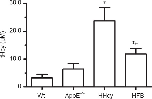
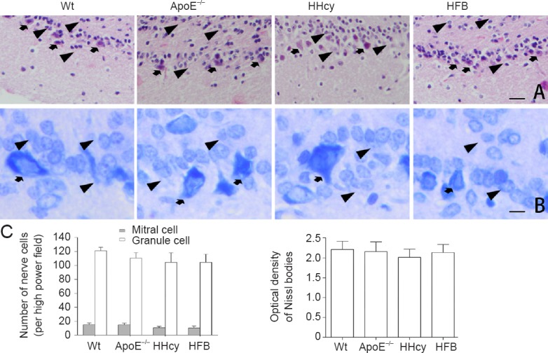
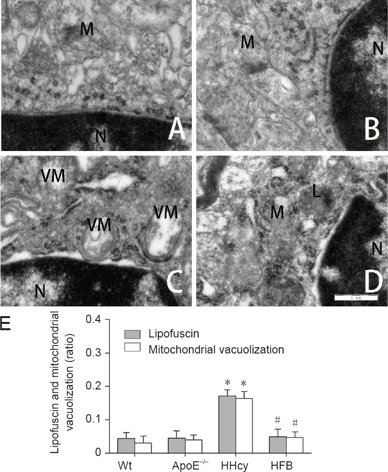
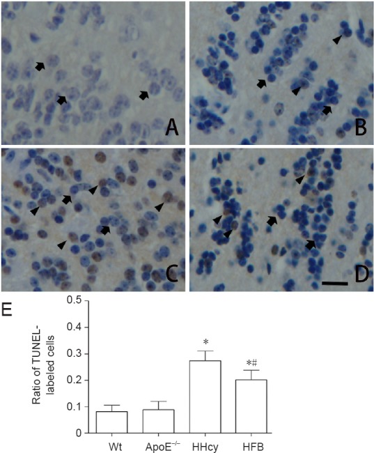
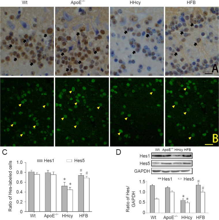
Hyperhomocysteinemia has been shown to be associated with neurodegenerative diseases; however, lesions or histological changes and mechanisms underlying homocysteine-induced injury in olfactory bulb neurons remain unclear. In this study, hyperhomocysteinemia was induced in apolipoprotein E-deficient mice with 1.7% methionine. Pathological changes in the olfactory bulb were observed through hematoxylin-eosin and Pischingert staining. Cell apoptosis in the olfactory bulb was determined through terminal deoxynucleotidyl transferase-mediated dUTP nick end-labeling (TUNEL) staining. Transmission electron microscopy revealed an abnormal ultrastructure of neurons. Furthermore, immunoreactivity and expression of the hairy enhancer of the split 1 (Hes1) and Hes5 were measured using immunohistochemistry, immunofluorescence, and western blot assay. Our results revealed no significant structural abnormality in the olfactory bulb of hyperhomocysteinemic mice. However, the number of TUNEL-positive cells increased in the olfactory bulb, lipofuscin and vacuolization were visible in mitochondria, and the expression of Hes1 and Hes5 decreased. These findings confirm that hyperhomocysteinemia induces injury in olfactory bulb neurons by downregulating Hes1 and Hes5 expression.
Keywords: Nissl body; apoptosis; hairy enhancer of split 1; hairy enhancer of split 5; homocysteine; nerve regeneration; neural regeneration; neurons; olfactory bulb.
Conflict of interest statement
None declared
Figures
Similar articles
-
Hyperhomocysteinemia-induced autophagy and apoptosis with downregulation of hairy enhancer of split 1/5 in cortical neurons in mice.Int J Immunopathol Pharmacol. 2017 Dec;30(4):371-382. doi: 10.1177/0394632017740061. Epub 2017 Nov 24. Int J Immunopathol Pharmacol. 2017. PMID: 29171783 Free PMC article.
-
Synaptic remodeling and reduced expression of the transcription factors, HES1 and HES5, in the cortex neurons of cognitively impaired hyperhomocysteinemic mice.Pathol Res Pract. 2020 Jun;216(6):152953. doi: 10.1016/j.prp.2020.152953. Epub 2020 Apr 9. Pathol Res Pract. 2020. PMID: 32345540
-
Cytoarchitecture of the olfactory bulb in the laggard mutant mouse.Neuroscience. 2014 Sep 5;275:259-71. doi: 10.1016/j.neuroscience.2014.06.011. Epub 2014 Jun 12. Neuroscience. 2014. PMID: 24931760
-
Hes1 and Hes5 as notch effectors in mammalian neuronal differentiation.EMBO J. 1999 Apr 15;18(8):2196-207. doi: 10.1093/emboj/18.8.2196. EMBO J. 1999. PMID: 10205173 Free PMC article.
-
Role of nerve growth factor in the olfactory system.Microsc Res Tech. 2002 Aug 1;58(3):197-203. doi: 10.1002/jemt.10149. Microsc Res Tech. 2002. PMID: 12203698 Review.
Cited by
-
Endogenous hydrogen sulfide and ERK1/2-STAT3 signaling pathway may participate in the association between homocysteine and hypertension.J Geriatr Cardiol. 2019 Nov;16(11):822-834. doi: 10.11909/j.issn.1671-5411.2019.11.007. J Geriatr Cardiol. 2019. PMID: 31853248 Free PMC article.
-
[High homocysteine level promotes autophagy and apoptosis of mouse hippocampal HT22 cells through the Notch1/Hes1 signaling pathway].Nan Fang Yi Ke Da Xue Xue Bao. 2023 Oct 20;43(10):1796-1803. doi: 10.12122/j.issn.1673-4254.2023.10.19. Nan Fang Yi Ke Da Xue Xue Bao. 2023. PMID: 37933657 Free PMC article. Chinese.
-
Neonatal-Inspired Reprogramming of Microglial Pan-Programmed Cell Death Enhances Regeneration in Adult Spinal Cord Injury.Research (Wash D C). 2025 Jul 2;8:0759. doi: 10.34133/research.0759. eCollection 2025. Research (Wash D C). 2025. PMID: 40607322 Free PMC article.
References
-
- Aléssio AC, Santos CX, Debbas V, Oliveira LC, Haddad R, Annichino-Bizzacchi JM. Evaluation of mild hyperhomocysteinemia during the development of atherosclerosis in apolipoprotein E-deficient and normal mice. Exp Mol Pathol. 2011;90:45–50. - PubMed
-
- Curro M, Gugliandolo A, Gangemi C, Risitano R, Ientile R, Caccamo D. Toxic effects of mildly elevated homocysteine concentrations in neuronal-like cells. Neurochem Res. 2014;39:1485–1495. - PubMed
LinkOut - more resources
Full Text Sources
Other Literature Sources
