

Whole-Brain Mapping of Direct Inputs to and Axonal Projections from GABAergic Neurons in the Parafacial Zone
- PMID: 29557546
- PMCID: PMC5960452
- DOI: 10.1007/s12264-018-0216-8
Whole-Brain Mapping of Direct Inputs to and Axonal Projections from GABAergic Neurons in the Parafacial Zone
Abstract
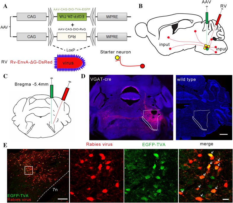
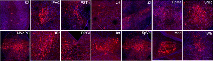
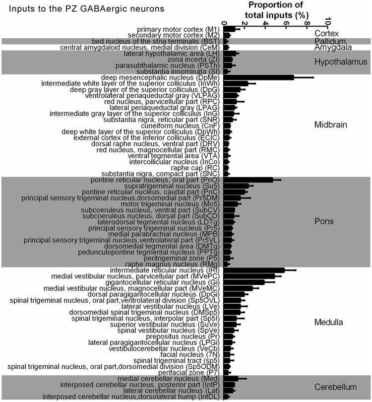
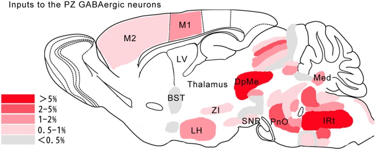
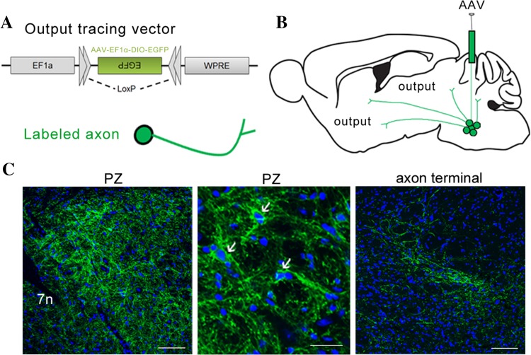
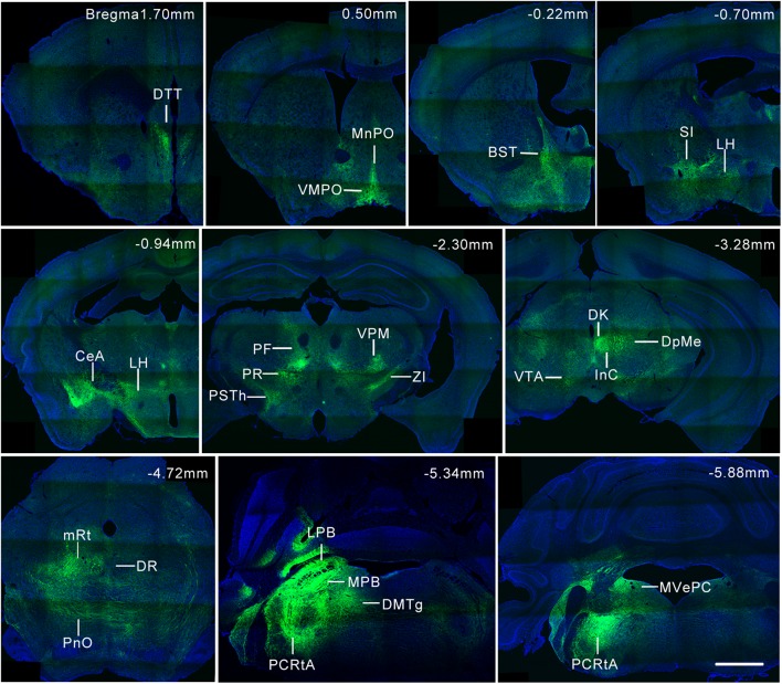
The GABAergic neurons in the parafacial zone (PZ) play an important role in sleep-wake regulation and have been identified as part of a sleep-promoting center in the brainstem, but the long-range connections mediating this function remain poorly characterized. Here, we performed whole-brain mapping of both the inputs and outputs of the GABAergic neurons in the PZ of the mouse brain. We used the modified rabies virus EnvA-ΔG-DsRed combined with a Cre/loxP gene-expression strategy to map the direct monosynaptic inputs to the GABAergic neurons in the PZ, and found that they receive inputs mainly from the hypothalamic area, zona incerta, and parasubthalamic nucleus in the hypothalamus; the substantia nigra, pars reticulata and deep mesencephalic nucleus in the midbrain; and the intermediate reticular nucleus and medial vestibular nucleus (parvocellular part) in the pons and medulla. We also mapped the axonal projections of the PZ GABAergic neurons with adeno-associated virus, and defined the reciprocal connections of the PZ GABAergic neurons with their input and output nuclei. The newly-found inputs and outputs of the PZ were also listed compared with the literature. This cell-type-specific neuronal whole-brain mapping of the PZ GABAergic neurons may reveal the circuits underlying various functions such as sleep-wake regulation.
Keywords: GABAergic neurons; Parafacial zone; Parvocellular reticular formation; Trans-synaptic tracing.
Conflict of interest statement
All authors claim that there are no conflicts of interest.
Figures
Similar articles
-
Whole-Brain Monosynaptic Inputs to Hypoglossal Motor Neurons in Mice.Neurosci Bull. 2020 Jun;36(6):585-597. doi: 10.1007/s12264-020-00468-9. Epub 2020 Feb 24. Neurosci Bull. 2020. PMID: 32096114 Free PMC article.
-
Forebrain GABAergic projections to locus coeruleus in mouse.J Comp Neurol. 2013 Jul 1;521(10):2373-97. doi: 10.1002/cne.23291. J Comp Neurol. 2013. PMID: 23296594 Free PMC article.
-
Whole-brain monosynaptic outputs and presynaptic inputs of GABAergic neurons in the vestibular nuclei complex of mice.Front Neurosci. 2022 Aug 26;16:982596. doi: 10.3389/fnins.2022.982596. eCollection 2022. Front Neurosci. 2022. PMID: 36090271 Free PMC article.
-
[Neurochemical mechanisms of sleep regulation].Glas Srp Akad Nauka Med. 2009;(50):97-109. Glas Srp Akad Nauka Med. 2009. PMID: 20666118 Review. Serbian.
-
Topographic and functional neuroanatomical study of GABAergic disinhibitory striatum-nigral inputs and inhibitory nigrocollicular pathways: neural hodology recruiting the substantia nigra, pars reticulata, for the modulation of the neural activity in the inferior colliculus involved with panic-like emotions.J Chem Neuroanat. 2006 Aug;32(1):1-27. doi: 10.1016/j.jchemneu.2006.05.002. Epub 2006 Jul 3. J Chem Neuroanat. 2006. PMID: 16820278 Review.
Cited by
-
Whole-Brain Monosynaptic Afferent Projections to the Cholecystokinin Neurons of the Suprachiasmatic Nucleus.Front Neurosci. 2018 Nov 5;12:807. doi: 10.3389/fnins.2018.00807. eCollection 2018. Front Neurosci. 2018. PMID: 30455627 Free PMC article.
-
A Whole-brain Map of Long-range Inputs to GABAergic Interneurons in the Mouse Caudal Forelimb Area.Neurosci Bull. 2020 May;36(5):493-505. doi: 10.1007/s12264-019-00458-6. Epub 2020 Jan 19. Neurosci Bull. 2020. PMID: 31956963 Free PMC article.
-
Coincident development and synchronization of sleep-dependent delta in the cortex and medulla.Curr Biol. 2024 Jun 17;34(12):2570-2579.e5. doi: 10.1016/j.cub.2024.04.064. Epub 2024 May 20. Curr Biol. 2024. PMID: 38772363 Free PMC article.
-
Rabies Virus Pseudotyped with CVS-N2C Glycoprotein as a Powerful Tool for Retrograde Neuronal Network Tracing.Neurosci Bull. 2020 Mar;36(3):202-216. doi: 10.1007/s12264-019-00423-3. Epub 2019 Aug 23. Neurosci Bull. 2020. PMID: 31444652 Free PMC article.
-
Morphological Tracing and Functional Identification of Monosynaptic Connections in the Brain: A Comprehensive Guide.Neurosci Bull. 2024 Sep;40(9):1364-1378. doi: 10.1007/s12264-024-01196-0. Epub 2024 May 3. Neurosci Bull. 2024. PMID: 38700806 Free PMC article. Review.
References
MeSH terms
Substances
LinkOut - more resources
Full Text Sources
Other Literature Sources
Miscellaneous
