. 2018 Oct 1;315(4):E594-E604.
doi: 10.1152/ajpendo.00343.2017. Epub 2018 Mar 20.

Fibroblast growth factor 23 does not directly influence skeletal muscle cell proliferation and differentiation or ex vivo muscle contractility

Affiliations

Fibroblast growth factor 23 does not directly influence skeletal muscle cell proliferation and differentiation or ex vivo muscle contractility

Keith G Avin et al. Am J Physiol Endocrinol Metab. .

Abstract

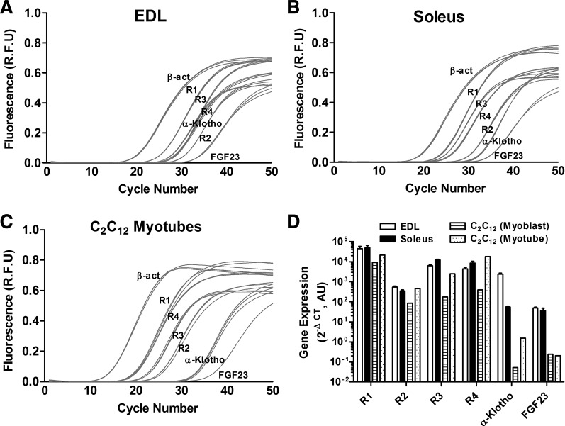
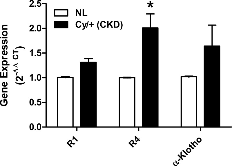
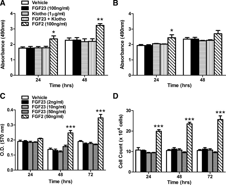
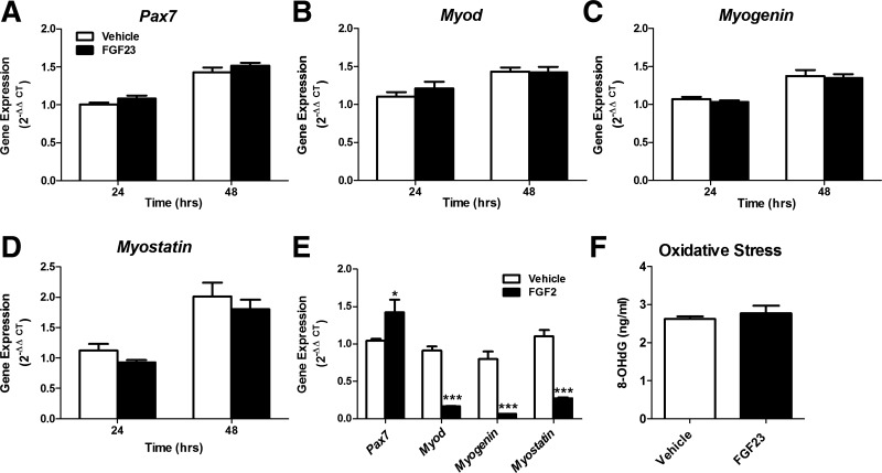
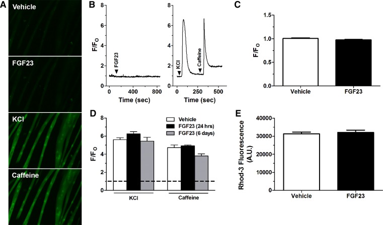
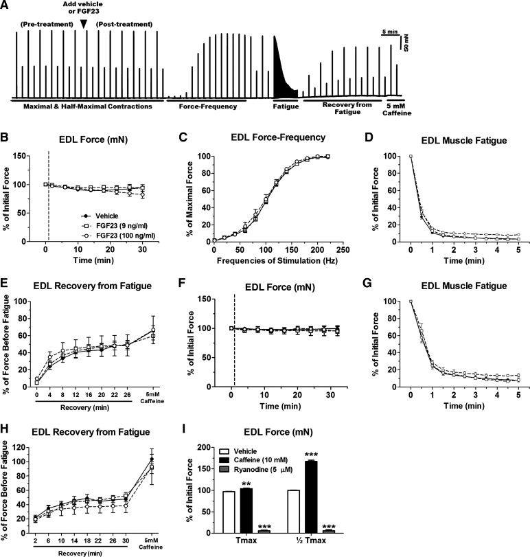
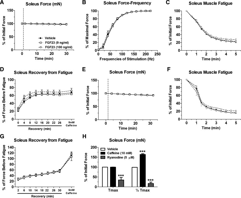
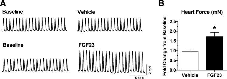
Skeletal muscle dysfunction accompanies the clinical disorders of chronic kidney disease (CKD) and hereditary hypophosphatemic rickets. In both disorders, fibroblast growth factor 23 (FGF23), a bone-derived hormone regulating phosphate and vitamin D metabolism, becomes chronically elevated. FGF23 has been shown to play a direct role in cardiac muscle dysfunction; however, it is unknown whether FGF23 signaling can also directly induce skeletal muscle dysfunction. We found expression of potential FGF23 receptors ( Fgfr1-4) and α-Klotho in muscles of two animal models (CD-1 and Cy/+ rat, a naturally occurring rat model of chronic kidney disease-mineral bone disorder) as well as C2C12 myoblasts and myotubes. C2C12 proliferation, myogenic gene expression, oxidative stress marker 8-OHdG, intracellular Ca2+ ([Ca2+]i), and ex vivo contractility of extensor digitorum longus (EDL) or soleus muscles were assessed after treatment with various amounts of FGF23. FGF23 (2-100 ng/ml) did not alter C2C12 proliferation, expression of myogenic genes, or oxidative stress after 24- to 72-h treatment. Acute or prolonged FGF23 treatment up to 6 days did not alter C2C12 [Ca2+]i handling, nor did acute treatment with FGF23 (9-100 ng/ml) affect EDL and soleus muscle contractility. In conclusion, although skeletal muscles express the receptors involved in FGF23-mediated signaling, in vitro FGF23 treatments failed to directly alter skeletal muscle development or function under the conditions tested. We hypothesize that other endogenous substances may be required to act in concert with FGF23 or apart from FGF23 to promote muscle dysfunction in hereditary hypophosphatemic rickets and CKD.

Keywords: chronic kidney disease; fibroblast growth factor 23; hypophosphatemic rickets; intracellular Ca2+; myogenesis.


Figures

Fig. 1.
Fig. 1.
Fibroblasti growth factor (Fgf) receptors (Fgfr), α-Klotho, and Fgf23 are expressed in skeletal muscle. AC: are representative real-time RT-PCR amplification plots showing values of fluorescence at each cycle number of Fgfr1–4, α-Klotho, and Fgf23 (each reaction in triplicate) in isolated extensor digitorum longus (EDL; A) and soleus (B) muscles from 4- to 5-mo-old CD-1 male mice and in differentiated C2C12 myotubes (C). D: summary data showing average 2ΔCT values (×106) of Fgfr1–4, α-Klotho, and Fgf23 in EDL and soleus muscles from 4- to 5-mo-old CD-1 male mice (n = 3) and in C2C12 myoblasts and myotubes (n = 1) expressed relative to β-actin on log10 scale.
Fig. 2.
Fig. 2.
Fgfr4 expression is elevated in the Cy/+ rat model for chronic kidney disease (CKD). Differences in gene expression of Fgfr1, r4, and α-Klotho in EDL muscle from Cy/+ rats or normal littermates (NL) were calculated using the 2ΔΔCT method (n = 6). *P < 0.05 vs. NL, 1-way ANOVA with Bonferroni post hoc analysis.
Fig. 3.
Fig. 3.
FGF23 treatment has no effect on C2C12 proliferation. A and B: cellular proliferation of undifferentiated C2C12 myoblasts (A) and differentiated C2C12 myotubes (B) treated with FGF23 (100 ng/ml), soluble Klotho (1 µg/ml), FGF23 + Klotho, or FGF2 (100 ng/ml, positive control) for 24 and 48 h, as assessed by absorbance at 490 nm using CellTiter 96 AQueous One kit (n = 3; 3 separate experiments). C: MTT assay of undifferentiated C2C12 myoblast proliferation after treatment with FGF23 (2–50 ng/ml) or FGF2 (50 ng/ml) for 24, 48, and 72 h (n = 4; repeated twice). D: hemocytometer-based cell count of undifferentiated C2C12 myoblasts after treatment with FGF23 (2–50 ng/ml) or FGF2 (50 ng/ml) for 24, 48, and 72 h (n = 3; repeated twice). *P < 0.05, **P < 0.01, and ***P < 0.001, 1-way ANOVA with Bonferroni post hoc analysis.
Fig. 4.
Fig. 4.
FGF23 treatment does not alter expression of myogenic markers or oxidative stress. AD: expression of myogenic gene markers Pax7 (A), Myod (B), Myogenin (C), and Myostatin (D) in differentiated C2C12 myotubes after treatment with FGF23 (100 ng/ml) or vehicle for 24 and 48 h. Gene expression was calculated using the 2ΔΔCT method (n = 6). E: positive control data showing expression of Pax7, Myod, Myogenin, and Myostatin in differentiated C2C12 myotubes after treatment with FGF2 (100 ng/ml) or vehicle for 48 h (n = 3). *P < 0.05 and ***P < 0.001, 1-way ANOVA with Bonferroni post hoc analysis. F: oxidative stress was assessed by measuring the DNA oxidative damage marker 8-hydroxy-2′-deoxyguanosine (8-OHdG) in differentiated C2C12 myotubes after 24-h treatment with FGF23 (100 ng/ml) or vehicle (n = 6). P > 0.05, Student’s t-test.
Fig. 5.
Fig. 5.
Neither acute nor long-term FGF23 treatment has an effect on C2C12 myotube [Ca2+]i. A: representative fluo-4 fluorescent images in C2C12 cells after vehicle, FGF23 (20 ng/ml), KCl (80 mM), or caffeine (20 mM) perfusion. Note the increase in fluorescence after KCl and caffeine treatment but not FGF23. B: representative fluo-4 fluorescence signal and perfusion protocol showing acute C2C12 myotube [Ca2+]i responses to FGF23 (20 ng/ml) (left) and KCl (80 mM) and caffeine (20 mM) perfusion (right). Arrowheads indicate points of perfusion of specific treatments. The fluorescence signal is expressed relative to baseline (F/FO). C: data summary of average peak change in fluo-4 fluorescence after perfusion with FGF23 (20 ng/ml) or vehicle expressed relative to baseline (F/FO) (n = 3 dishes/group with 7–8 myotubes/dish averaged). P > 0.05, Student’s t-test. D: average change in fluo-4 fluorescence in response to perfusion with KCl (80 mM) and caffeine (20 mM) in myotubes incubated with FGF23 (20 ng/ml) for 24 h or 6 days (n = 3–6 dishes/group with 5–11 myotubes/dish averaged). Horizontal dashed line indicates the fluorescence level at baseline before stimulation of Ca2+ release. P > 0.05, 1-way ANOVA. E: Rhod-3 fluorescence in differentiated C2C12 myocytes after treatment with FGF23 (100 ng/ml) for 4 h (n = 6). P > 0.05, Student’s t-test.
Fig. 6.
Fig. 6.
Acute FGF23 administration does not alter CD-1 mouse EDL muscle contractile properties. A: representative ex vivo contractility force data obtained from 1 muscle (x-axis: time; y-axis: force) showing an entire contraction protocol and time of FGF23 addition. B: maximal tetanic force output (at 200-Hz stimulation) from CD-1 mouse EDL muscles after treatment with vehicle or FGF23 expressed relative to values before vehicle or FGF23 application. C: force-frequency relationship of vehicle- or FGF23-treated EDL muscles stimulated to contract with frequencies in the range of 1–220 Hz. Forces at each frequency are expressed relative to the maximal force obtained. D: time course of maximal tetanic force decline during a fatiguing protocol in vehicle- or FGF23-treated EDL muscles. Force at each time point is expressed relative to force just before fatigue. E: maximal tetanic force recovery during various time points postfatigue and with the addition of 5 mM caffeine in vehicle- or FGF23-treated EDL muscles. F: half-maximal tetanic force output (at 100-Hz stimulation) from CD-1 mouse EDL muscles after treatment with vehicle or FGF23 expressed relative to values before vehicle or FGF23 application. G: time course of half-maximal tetanic force decline during a fatiguing protocol in vehicle- or FGF23-treated EDL muscles. Force at each time point is expressed relative to force just before fatigue. H: half-maximal tetanic force recovery during various time points postfatigue and with the addition of 5 mM caffeine in vehicle- or FGF23-treated EDL muscles. I: maximal and half-maximal tetanic force after exposure of EDL muscles to 10 mM caffeine or 5 µM ryanodine, as positive controls, expressed as %force before vehicle or FGF23 treatment (however, error bars that are present in each group may be too small to be seen). Dashed lines in B and F represent points at which FGF23 was added to the muscle contractility bath; n = 3–10 muscles/group. **P < 0.01 and ***P < 0.001, 2-way ANOVA with Bonferroni post hoc analysis.
Fig. 7.
Fig. 7.
Acute FGF23 administration does not alter CD-1 mouse soleus muscle contractile properties. A: maximal tetanic force output (at 140–160 Hz stimulation) from CD-1 mouse soleus muscles after treatment with vehicle or FGF23 expressed relative to values before vehicle or FGF23 application. B: force-frequency relationship of vehicle- or FGF23-treated soleus muscles stimulated to contract with frequencies in the range of 1–220 Hz. Forces at each frequency are expressed relative to the maximal force obtained. C: time course of maximal tetanic force decline during a fatiguing protocol in vehicle- or FGF23-treated soleus muscles. Force at each time point is expressed relative to force just before fatigue. D: maximal tetanic force recovery during various time points postfatigue and with the addition of 5 mM caffeine in vehicle- or FGF23-treated soleus muscles. E: half-maximal tetanic force output (at 40 Hz stimulation) after treatment with vehicle or FGF23 expressed relative to values before vehicle or FGF23 application. F: time course of half-maximal tetanic force decline during a fatiguing protocol in vehicle- or FGF23-treated soleus muscles. Force at each time point is expressed relative to force just before fatigue. G: half-maximal tetanic force recovery during various time points postfatigue and with the addition of 5 mM caffeine in vehicle- or FGF23-treated soleus muscles. H: maximal and half-maximal tetanic force after exposure of soleus muscles to 10 mM caffeine or 5 µM ryanodine, as positive controls, expressed as a %force before vehicle or FGF23 treatment (however, error bars that are present in each group may be too small to be seen). Dashed lines in A and E represent points at which FGF23 was added to the muscle contractility bath; n = 5–10 muscles/group. ***P < 0.001, 2way ANOVA with Bonferroni post hoc analysis.
Fig. 8.
Fig. 8.
Acute FGF23 administration increases isolated heart contractility. A: representative force tracings of isolated mouse heart muscle paced at 1 Hz at baseline (before vehicle or FGF23 treatment) and after 30 min of exposure to vehicle or 9 ng/ml FGF23. B: summary data of whole heart contractile force output after 30 min of vehicle or 9 ng/ml FGF23 exposure. Data are expressed as fold change of contractile force relative to baseline; n = 4–6 hearts/group. *P < 0.05, 2-tailed t-test.

References

    1. Abboud M, Rybchyn MS, Liu J, Ning Y, Gordon-Thomson C, Brennan-Speranza TC, Cole L, Greenfield H, Fraser DR, Mason RS. The effect of parathyroid hormone on the uptake and retention of 25-hydroxyvitamin D in skeletal muscle cells. J Steroid Biochem Mol Biol 173: 173–179, 2017. doi:10.1016/j.jsbmb.2017.01.001. - DOI - PubMed
    1. Adams GR, Vaziri ND. Skeletal muscle dysfunction in chronic renal failure: effects of exercise. Am J Physiol Renal Physiol 290: F753–F761, 2006. doi:10.1152/ajprenal.00296.2005. - DOI - PubMed
    1. Allen DG, Westerblad H. The effects of caffeine on intracellular calcium, force and the rate of relaxation of mouse skeletal muscle. J Physiol 487: 331–342, 1995. doi:10.1113/jphysiol.1995.sp020883. - DOI - PMC - PubMed
    1. Allen RE, Boxhorn LK. Regulation of skeletal muscle satellite cell proliferation and differentiation by transforming growth factor-beta, insulin-like growth factor I, and fibroblast growth factor. J Cell Physiol 138: 311–315, 1989. doi:10.1002/jcp.1041380213. - DOI - PubMed
    1. Aono Y, Hasegawa H, Yamazaki Y, Shimada T, Fujita T, Yamashita T, Fukumoto S. Anti-FGF-23 neutralizing antibodies ameliorate muscle weakness and decreased spontaneous movement of Hyp mice. J Bone Miner Res 26: 803–810, 2011. doi:10.1002/jbmr.275. - DOI - PubMed

Publication types

MeSH terms

LinkOut - more resources