

Optimal model-based sensorless adaptive optics for epifluorescence microscopy
- PMID: 29558510
- PMCID: PMC5860766
- DOI: 10.1371/journal.pone.0194523
Optimal model-based sensorless adaptive optics for epifluorescence microscopy
Abstract
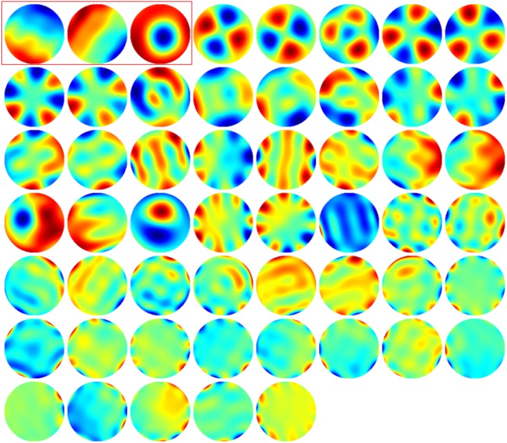
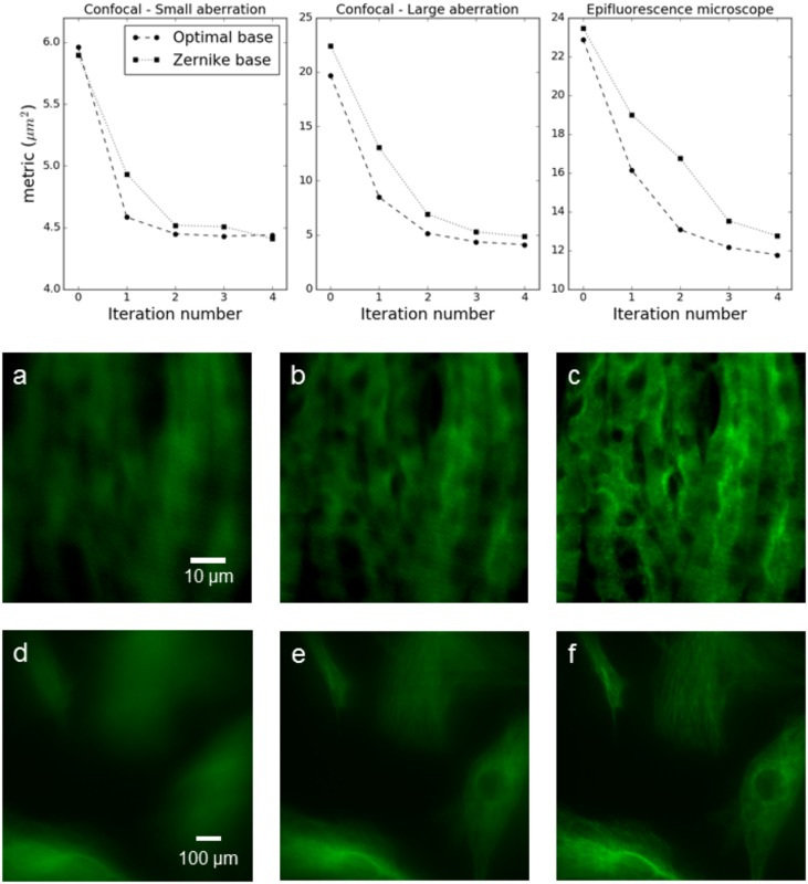
We report on a universal sample-independent sensorless adaptive optics method, based on modal optimization of the second moment of the fluorescence emission from a point-like excitation. Our method employs a sample-independent precalibration, performed only once for the particular system, to establish the direct relation between the image quality and the aberration. The method is potentially applicable to any form of microscopy with epifluorescence detection, including the practically important case of incoherent fluorescence emission from a three dimensional object, through minor hardware modifications. We have applied the technique successfully to a widefield epifluorescence microscope and to a multiaperture confocal microscope.
Conflict of interest statement
Figures


Similar articles
-
Aberration correction during real time in vivo imaging of bone marrow with sensorless adaptive optics confocal microscope.J Biomed Opt. 2014 Aug;19(8):086009. doi: 10.1117/1.JBO.19.8.086009. J Biomed Opt. 2014. PMID: 25117079
-
Adaptive aberration correction in a confocal microscope.Proc Natl Acad Sci U S A. 2002 Apr 30;99(9):5788-92. doi: 10.1073/pnas.082544799. Epub 2002 Apr 16. Proc Natl Acad Sci U S A. 2002. PMID: 11959908 Free PMC article.
-
Sensorless adaptive optics implementation in widefield optical sectioning microscopy inside in vivo Drosophila brain.J Biomed Opt. 2016 Mar;21(3):36006. doi: 10.1117/1.JBO.21.3.036006. J Biomed Opt. 2016. PMID: 26968001
-
Digital imaging microscopy of living cells.Trends Cell Biol. 1998 Jul;8(7):288-92. doi: 10.1016/s0962-8924(98)01301-4. Trends Cell Biol. 1998. PMID: 9714601 Review.
-
Two-photon excitation imaging based on a compact scanning head.IEEE Eng Med Biol Mag. 1999 Sep-Oct;18(5):18-22, 30. doi: 10.1109/51.790987. IEEE Eng Med Biol Mag. 1999. PMID: 10497738 Review. No abstract available.
Cited by
-
Model reference adaptive system and pseudo-sliding mode control with exponential reaching law for sensorless-speed control of PMSM.PLoS One. 2025 May 19;20(5):e0321985. doi: 10.1371/journal.pone.0321985. eCollection 2025. PLoS One. 2025. PMID: 40388518 Free PMC article.
References
-
- Débarre D, Botcherby EJ, Booth MJ, Wilson T. Adaptive optics for structured illumination microscopy. Optics express, 2008; 16(13): 9290–9305. doi: 10.1364/OE.16.009290 - DOI - PubMed
-
- Débarre D, Botcherby EJ, Watanabe T, Srinivas S, Booth MJ, Wilson T. Image-based adaptive optics for two-photon microscopy. Optics letters, 2009; 34(16): 2495–2497. doi: 10.1364/OL.34.002495 - DOI - PMC - PubMed
-
- Bourgenot C, Taylor JM, Saunter CD, Girkin JM, and Love GD. AO modal optimization in a live, beating zebrafish heart. Adaptive Optics: Methods, Analysis and Applications. Optical Society of America, 2013. p. OW4A. 4.
-
- Facomprez A, Beaurepaire E, Débarre D. Accuracy of correction in modal sensorless adaptive optics. Optics express, 2012; 20(3): 2598–2612. doi: 10.1364/OE.20.002598 - DOI - PubMed
-
- Thayil A, Booth MJ. Self calibration of sensorless adaptive optical microscopes. Journal of the European Optical Society-Rapid publications, 2011; 6.
Publication types
MeSH terms
LinkOut - more resources
Full Text Sources
Other Literature Sources
