A white membrane beneath the inner limiting membrane of the retina in a 4-year-old child with ultrastructural evidence: a case report
- PMID: 29558906
- PMCID: PMC5859535
- DOI: 10.1186/s12886-018-0748-8
A white membrane beneath the inner limiting membrane of the retina in a 4-year-old child with ultrastructural evidence: a case report
Abstract
Background: Epiretinal membranes (ERMs), secondary to retinal cell proliferation on the retinal surface, usually affect patients over 50 years of age but occur rarely in children. Here we report the case of a 4-year-old patient with a unilateral sub-inner limiting membrane (sub-ILM) membrane mimicking epiretinal membrane with notable ultrastructural features indicating its possible origin from old sub-ILM hemorrhage.
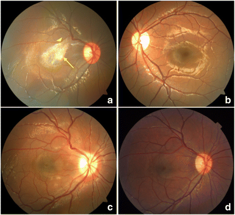
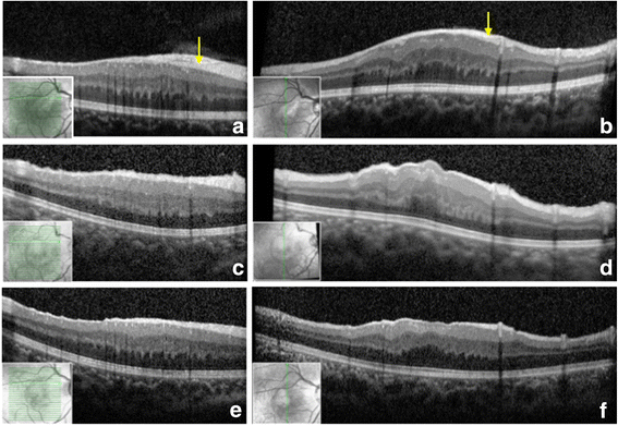
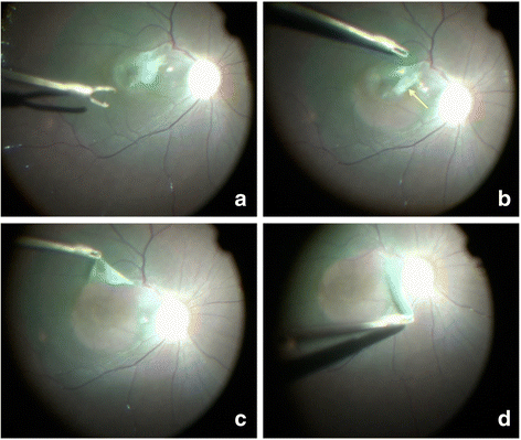
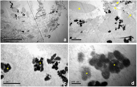
Case presentation: A 4-year-old boy was admitted with the complaint of poor vision in his right eye, which had been detected at school vision screening performed 6 months earlier. Fundal examination showed a feather-shaped white membrane in the macula of the right eye, and optical coherence tomography (OCT) revealed a thickened retina with a hyper-reflective band on the retinal nerve fiber layer. We suspected epiretinal membrane in the right eye, and pars plana vitrectomy with membrane peeling was performed to improve the patient's vision. Surprisingly, the membrane was found intraoperatively to be located beneath the intact ILM; it was lifted carefully from the underlying retina as it was strongly adhered to a retinal artery of the superotemporal arcade. Postoperative scanning electron microscopy showed that the membrane consisted of hemosiderin, collagenous fibre and fibrinoid deposits. At follow-up visits, fundal examination and OCT revealed improvement in the retinal structure with disappearance of the hyper-reflective band and reduced retinal thickness. The patient's visual acuity in the right eye was stable at 20/100 at 1 year post operation.
Conclusions: The white membrane presented here was found to lie between the intact ILM and the rest of the retina, adhering firmly to the superotemporal vessel arch. Given the ultrastructural findings of the membrane and the medical history, we speculate that the sub-ILM membrane probably developed secondary to a sub-ILM hemorrhage.
Keywords: Case report; Children; Sub-inner limiting membrane hemorrhage; Sub-inner limiting membrane membrane; Ultrastuctural pathology; Vitrectomy.
Conflict of interest statement
Ethics approval and consent to participate
The study was approved by the Ethics Committee of the Eye and Ear Nose Throat Hospital of Fudan University, and the procedures conformed to the tenets of the Declaration of Helsinki.
Consent for publication
Written informed consent for the publication of the case report and the accompanying images has been obtained from the patient’s father.
Competing interests
The authors declare that they have no competing interests.
Publisher’s Note
Springer Nature remains neutral with regard to jurisdictional claims in published maps and institutional affiliations.
Figures


Similar articles
-
Ultrastructural changes of the vitreoretinal interface during long-term follow-up after removal of the internal limiting membrane.Am J Ophthalmol. 2014 Sep;158(3):550-6.e1. doi: 10.1016/j.ajo.2014.05.022. Epub 2014 May 27. Am J Ophthalmol. 2014. PMID: 24878309
-
Value of internal limiting membrane peeling in surgery for idiopathic macular hole and the correlation between function and retinal morphology.Acta Ophthalmol. 2009 Dec;87 Thesis 2:1-23. doi: 10.1111/j.1755-3768.2009.01777.x. Acta Ophthalmol. 2009. PMID: 19912135 Clinical Trial.
-
The role of membrane-inner retina adherence in predicting simultaneous internal limiting membrane peeling during idiopathic epiretinal membrane surgery.Eye (Lond). 2017 Apr;31(4):636-642. doi: 10.1038/eye.2016.285. Epub 2016 Dec 23. Eye (Lond). 2017. PMID: 28009345 Free PMC article.
-
Early and late inner retinal changes after inner limiting membrane peeling.Int Ophthalmol. 2014 Apr;34(2):437-46. doi: 10.1007/s10792-013-9831-6. Epub 2013 Jul 18. Int Ophthalmol. 2014. PMID: 23864285 Review.
-
THE EFFECT OF INTERNAL LIMITING MEMBRANE PEELING ON IDIOPATHIC EPIRETINAL MEMBRANE SURGERY, WITH A REVIEW OF THE LITERATURE.Retina. 2017 May;37(5):873-880. doi: 10.1097/IAE.0000000000001263. Retina. 2017. PMID: 27617536 Review.
Cited by
-
Pars plana vitrectomy with intraoperative optical coherence tomography for sub-internal limiting membrane fibrosis excision in a child with Terson syndrome: Surgical and pathological correlation.Am J Ophthalmol Case Rep. 2019 May 31;15:100479. doi: 10.1016/j.ajoc.2019.100479. eCollection 2019 Sep. Am J Ophthalmol Case Rep. 2019. PMID: 31198885 Free PMC article.
References
Publication types
MeSH terms
Substances
Grants and funding
LinkOut - more resources
Full Text Sources
Other Literature Sources
Medical
Miscellaneous

