Review
. 2018 Mar 20;22(1):75.
doi: 10.1186/s13054-018-1997-x.

Continuous Electroencephalography Monitoring in Adults in the Intensive Care Unit

Affiliations
Review

Continuous Electroencephalography Monitoring in Adults in the Intensive Care Unit

Anselmo Caricato et al. Crit Care. .

Abstract

This article is one of ten reviews selected from the Annual Update in Intensive Care and Emergency Medicine 2018. Other selected articles can be found online at https://www.biomedcentral.com/collections/annualupdate2018 . Further information about the Annual Update in Intensive Care and Emergency Medicine is available from http://www.springer.com/series/8901 .


Conflict of interest statement

Ethics approval and consent to participate

Not applicable

Consent for publication

Not applicable

Competing interests

The authors declare that they have no competing interests.

Publisher’s Note

Springer Nature remains neutral with regard to jurisdictional claims in published maps and institutional affiliations.

Figures
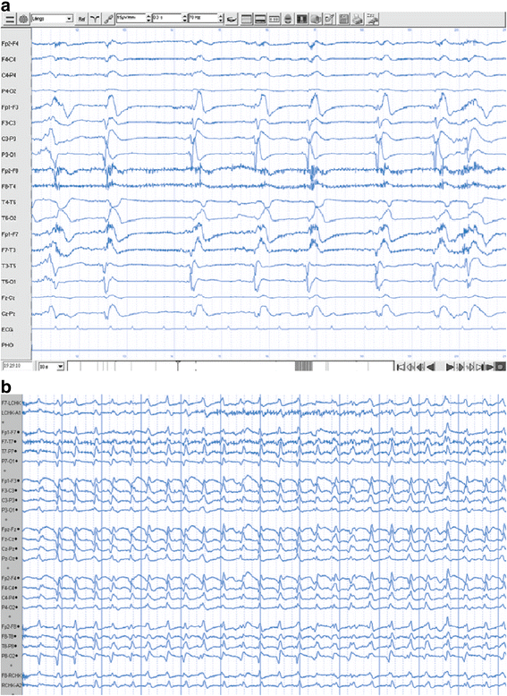
Fig. 1
Fig. 1
Lateralized periodic discharges and generalized periodic discharges. An example of two periodic patterns: a temporo-parietal lateralized periodic discharges and b generalized periodic discharges. Continuous EEG recording is recommended in these cases
Fig. 2
Fig. 2
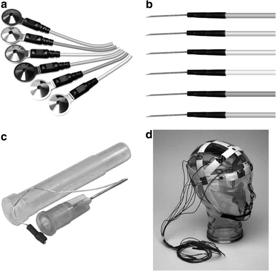
Type of EEG electrodes. Examples of classic cup electrodes (a), subdermal needle electrodes (b), subdermal wire electrodes (c) and an EEG template system (d)
Fig. 3
Fig. 3
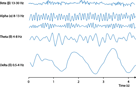
EEG waveform frequency. Examples of traditional classification of alpha, beta, theta, delta EEG frequencies
Fig. 4
Fig. 4
Color density spectral array. In this frequency(y) vs time(x) graph, each pixel shows the amplitude of EEG recording, according to its color (purple low, red high). The color range for the amplitude scale is shown on the left. Waveform frequency is displayed on the right. Phases of sudden reduction of faster frequencies during sedation can be easily recognized (arrows)
Fig. 5
Fig. 5
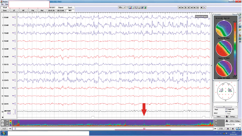
Focal increase in EEG fast frequencies. In this raw EEG, fast frequencies in the left temporal region are shown. By DSA compression, these are easily observed as a sudden modification in the EEG recording (arrow). Topographic maps of spectral power are displayed on the right; they are spatial representations of the amplitude of traditional waveform frequencies (delta, theta, alpha), referred to the raw EEG displayed on the screen. Focal asymmetries can be easily identified. The color range of the amplitude scale is shown on the right

References

    1. Nuwer MR. EEG and evoked potentials: monitoring cerebral function in the neurosurgical ICU. In: Martin NA, editor. Neurosurgical Intensive Care. Philadelphia: W.B. Saunders; 1994. pp. 647–659. - PubMed
    1. Ney JP, van der Goes DN, Nuwer MR, Nelson L, Eccher MA. Continuous and routine EEG in intensive care: utilization and outcomes, United States 2005–2009. Neurology. 2013;81:2002–2008. doi: 10.1212/01.wnl.0000436948.93399.2a. - DOI - PMC - PubMed
    1. Kinney MO, Kaplan PW. An update on the recognition and treatment of non-convulsive status epilepticus in the intensive care unit. Expert Rev Neurother. 2017;17:987–1002. doi: 10.1080/14737175.2017.1369880. - DOI - PubMed
    1. Herman ST, Abend N, Bleck TP. Consensus statement on continuous eeg in critically ill adults and children, Part I: Indications. J Clin Neurophysiol. 2015;32:87–95. doi: 10.1097/WNP.0000000000000166. - DOI - PMC - PubMed
    1. Claassen J, Taccone FS, Horn P, Holtkamp M, Stocchetti N, Oddo M. Recommendations on the use of EEG monitoring in critically ill patients: consensus statement from the neurointensive care section of the ESICM. Intensive Care Med. 2013;39:1337–1351. doi: 10.1007/s00134-013-2938-4. - DOI - PubMed

LinkOut - more resources