. 2018 Mar 20;18(1):104.
doi: 10.1186/s12906-018-2165-1.

Gelidiella acerosa inhibits lung cancer proliferation

Affiliations

Gelidiella acerosa inhibits lung cancer proliferation

Fazeela Mahaboob Begum S M et al. BMC Complement Altern Med. .

Abstract

Background: Lung adenocarcinoma is the most common subtype of Non small cell lung cancer in which the PI3K/Akt cascade is frequently deregulated. The ubiquitous expression of the PI3K and the frequent inactivation of PTEN accounts for the prolonged survival, evasion of apoptosis and metastasis in cancer. This has led to the development of PI3K inhibitors in the treatment of cancer. Synthetic PI3K inhibitors undergoing clinical and preclinical studies are toxic in animals. Hence, there is a critical need to identify PI3K inhibitor(s) of natural origin. The current study aims to explore the efficacy of the red algae Gelidiella acerosaon inhibition of cell proliferation, migration and the expression of cell survival genes in lung adenocarcinoma cell line A549.

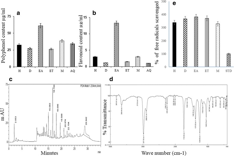
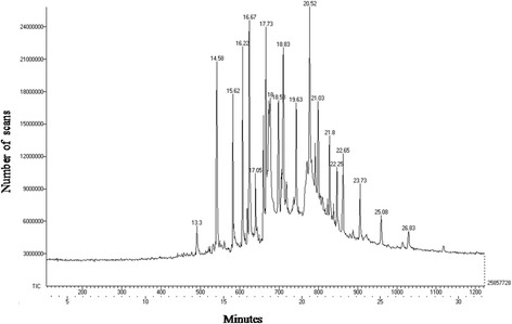
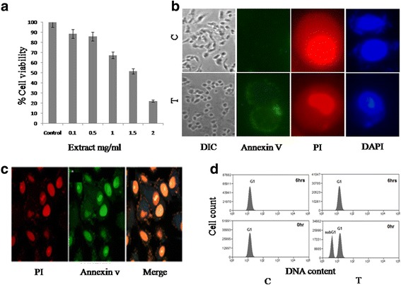
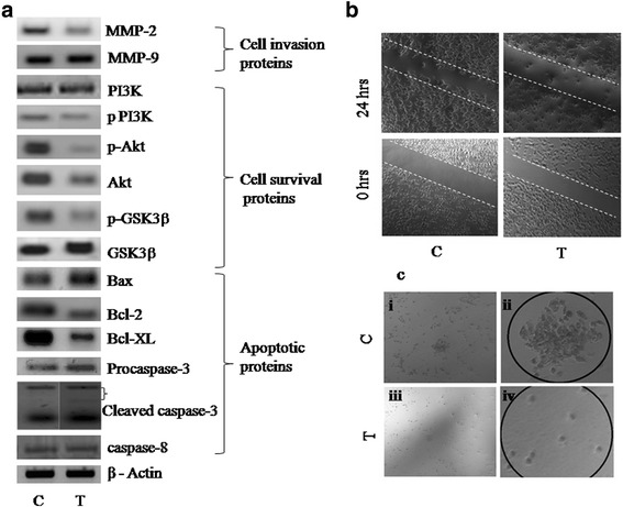
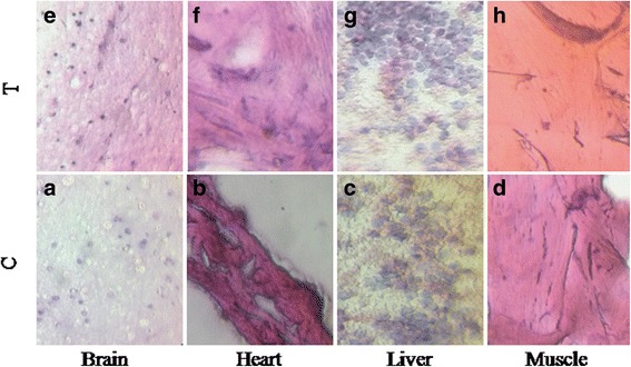
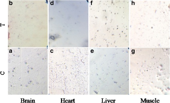
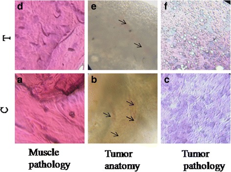
Methods: The phytoconstituents of Gelidiella acerosa were extracted sequentially with solvents of different polarity, screened qualitatively and quantitatively for secondary metabolites and characterized by GC-MS. The in-vitro studies were performed to check the efficacy of the extract on cell proliferation (MTT assay), cell invasion (scratch assay and colony formation assay), apoptosis (fluorescent, confocal microscopy and flow cytometry) and expression of apoptosis and cell survival proteins including PI3K, Akt and GSK3β and matrix metalloproteinase MMP2 and MMP9 by Western blot method. The antitumor activity of GAE was analyzed in a tumor model of Zebrafish.

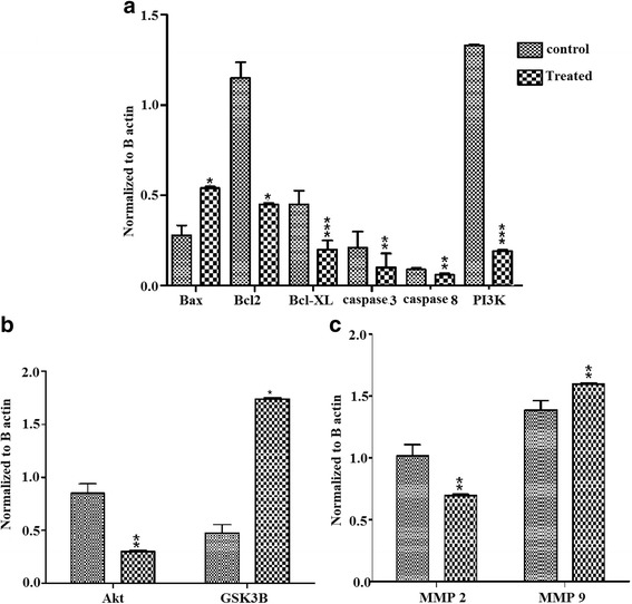
Results: The outcomes of the in vitro analysis showed an inhibition of cell proliferation, induction of apoptosis, inhibition of cell migration and colonization by the crude extract. The analysis of protein expression showed the activation of caspases 3 and Pro apoptotic protein Bax accompanied by decreased expression of Bcl-2 and Bcl-XL. On the other hand, subsequent activation of GSK3β and down regulation of PI3K, Akt were observed. The decreased expression of MMP2 correlated with the antimetastatic activity of the extract. The in vivo studies showed an inhibition of tumor growth by GAE in Zebrafish.

Conclusion: The phytoconstituents of algal extract contributed to the anticancer properties as evidenced by in vitro and in vivo studies. These phytoconstituents can be considered as a natural source of PI3K/Akt inhibitor for treatment of cancers involving the PI3K cascade.

Keywords: Akt; Bax; Bcl-2; Bcl-XL; Caspase 3; GSK3β; Gelidiella acerosa; PI3K; Tumor model of zebrafish.


Conflict of interest statement

Ethics approval

The Ethical clearance was granted by the Institutional Animal Ethics Committee of Pentagrit, where the work was carried out (IAEC study No: 213/GO06/IAEC).

Consent for publication

Not applicable.

Competing interests

The authors declare that they have no competing interests.

Publisher’s Note

Springer Nature remains neutral with regard to jurisdictional claims in published maps and institutional affiliations.

Figures

Fig. 1
Fig. 1
Characterization of algal phytoconstituents. a and b Quantification of polyphenols and flavonoids in algal extracts reveals maximum concentration in ethyl acetate extract. c. HPLC analysis of ethyl acetate extract shows the presence of 4 compounds with retention time. d FTIR analysis of ethyl acetate extract showing presence of functional groups. e Analysis of antioxidant activity by DPPH assay reveals maximum antioxidant efficiency of ethyl acetate extract. Values expressed are Mean ± S.D. (H- hexane, D- Dichloromethane, EA- ethyl acetate, ET- ethanol, M- methanol, AQ - aqueous, STD- Ascorbic acid)
Fig. 2
Fig. 2
GC-MS profile of GAE. The GC-MS analysis revealed the presence of various compounds with differing retention time
Fig. 3
Fig. 3
In vitro analysis of anticancer activity in A549 cells. a Cytotoxicity analysis in A549 cells identifies IC50 as1.5 mg/ml of GAE. b Fluorescent imaging with Annexin, Propidium iodide and DAPI revealed the translocation of Phosphatidyl serine to the cell membrane and nuclear fragmentation in GAE treated cells. c Confocal imaging of GAE treated cells showing nuclear fragmentation. d FACS analysis of Apoptosis by DAPI staining shows the emergence of apoptotic peak after 6 h of GAE treatment. All experiments were done in triplicate. The values represent Mean ± SD. C-control, T-treated
Fig. 4
Fig. 4
Analysis of protein expression. a Western blot analysis of procaspase-3, cleaved caspase3, caspase-8, Bax, Bcl-2 and Bcl-XL, p-Akt, Akt, p-GSK3β and GSK3β, PI3K, p-PI3K, MMP2 and MMP9. βactin was used to check equal loading. b The scratch assay shows the inhibition of cell invasion into the wound region after 24 h of GAE treatment (1 mg/ml). c The clonogenic assay result shows the inability of single cells to proliferate as a colony after GAE treatment (1 mg/ml). (i) Colony formed in control cells after 7 days, (ii) magnified colony of control cells, (iii) Treated cells after 7 days, (iv) treated cells magnified. C-control, T-treated
Fig. 5
Fig. 5
Analysis of protein expression. Histogram shows the optical density of (a) Bax, Bcl2, Bcl-XL, Caspase 8, Caspase 3 and ratio of phospho to total PI3K. b Histogram shows the optical density of phospho to total Akt, GSK3β. c Histogram shows the optical density of MMP2 and MMP9. Relative expression of protein was normalized to β actin. The density was analyzed by ImageJ software. Values represent mean ± SD.*p < 0.1, **p < 0.05, ***p < 0.001 compared to control group. C-control, T-treated
Fig. 6
Fig. 6
In vivo chronic toxicology analysis. a, e Shows the unaltered brain pathology of control and GAE treated Zebrafish. b, f Shows the pathology of heart muscles in control and GAE treated Zebrafish. c, g Shows the unaltered muscle pathology of control and GAE treated Zebrafish. d, h Shows the pathology of liver cells in control and GAE treated Zebrafish. The pathology results confirm that GAE is not toxic to these major organs under chronic conditions. C-control, T-treated
Fig. 7
Fig. 7
Toxicology analysis in Tissue-Chip. The pathology studies of vital organs namely brain, heart, liver and muscle were analyzed in both control (a, c, e, g) and in treated (b, d, f, h) tissue chip. The treatment with algal extract 500 μg did not affect the tissue architecture. The results of tissue chip analysis also support GAE to be safe without toxicity. These tissues were developed and analysed for toxicity in Tissue-Chip Pentagrit, Chennai. C-control, T-treated
Fig. 8
Fig. 8
In vivo analysis of antitumor activity. a, b, c Histopathological analysis of muscle and tumor of tumor induced zebrafish. d, e, f Histopathological analysis of muscle and tumors from GAE treated tumor induced zebrafish. C-control, T-treated

References

    1. Kumar R. A phase 1b trail of the combination of an all- oral regimen of capecitabine and erlotinib in advanced non small cell lung cancer in Caucasian patients. Cancer ChemotherPharmacol. 2016;77:375–383. - PubMed
    1. Travis WD, Brambilla E, Noguchi M, Nicholson AG, Geisinger KR, Yatabe Y, et al. International association for the study of lung cancer/american thoracic society/european respiratory society: international multidisciplinary classification of lung adenocarcinoma. J Thorac Oncol. 2011;8(2):381–385. - PubMed
    1. Dacic S. Molecular diagnostics of lung carcinomas. Arch Pathol Lab Med. 2011;135(5):622–629. - PubMed
    1. RapariaK VC, DeCamp MM, Patel JD, Mehta MP. Molecular profiling in non–small cell lung cancer :a step toward personalized medicine. Arch Pathol Lab Med. 2013;137:481–491. doi: 10.5858/arpa.2012-0287-RA. - DOI - PubMed
    1. Fruman DA, Meyers RE, Cantley LC. Phosphoinositide kinases. Annu Rev Biochem. 1998;67:481–507. doi: 10.1146/annurev.biochem.67.1.481. - DOI - PubMed

Substances

LinkOut - more resources