

Diagnostic value of cerebrospinal fluid tau, neurofilament, and progranulin in definite frontotemporal lobar degeneration
- PMID: 29559004
- PMCID: PMC5859717
- DOI: 10.1186/s13195-018-0364-0
Diagnostic value of cerebrospinal fluid tau, neurofilament, and progranulin in definite frontotemporal lobar degeneration
Abstract
Background: We explored the diagnostic performance of cerebrospinal fluid (CSF) biomarkers in allowing differentiation between frontotemporal lobar degeneration (FTLD) and Alzheimer's disease (AD), as well as between FTLD pathological subtypes.
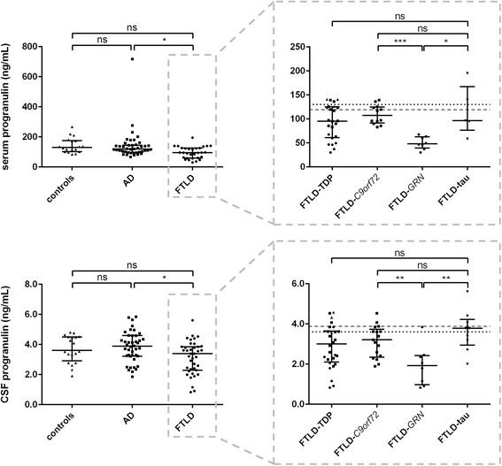
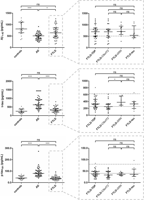
Methods: CSF levels of routine AD biomarkers (phosphorylated tau (p-tau181), total tau (t-tau), and amyloid-beta (Aβ)1-42) and neurofilament proteins, as well as progranulin levels in both CSF and serum were quantified in definite FTLD (n = 46), clinical AD (n = 45), and cognitively healthy controls (n = 20). FTLD subgroups were defined by genetic carrier status and/or postmortem neuropathological confirmation (FTLD-TDP: n = 34, including FTLD-C9orf72: n = 19 and FTLD-GRN: n = 9; FTLD-tau: n = 10).
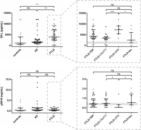
Results: GRN mutation carriers had significantly lower progranulin levels compared to other FTLD patients, AD, and controls. Both t-tau and p-tau181 were normal in FTLD patients, even in FTLD-tau. Aβ1-42 levels were very variable in FTLD. Neurofilament light chain (Nf-L) was significantly higher in FTLD compared with AD and controls. The reference logistic regression model based on the established AD biomarkers could be improved by the inclusion of CSF Nf-L, which was also important for the differentiation between FTLD and controls. Within the FTLD cohort, no significant differences were found between FTLD-TDP and FTLD-tau, but GRN mutation carriers had higher t-tau and Nf-L levels than C9orf72 mutation carriers and FTLD-tau patients.
Conclusions: There is an added value for Nf-L in the differential diagnosis of FTLD. Progranulin levels in CSF depend on mutation status, and GRN mutation carriers seem to be affected by more severe neurodegeneration.
Keywords: Alzheimer’s disease; Biomarkers; Cerebrospinal fluid; Differential diagnosis; Frontotemporal lobar degeneration; Neurofilament; Progranulin; Tau.
Conflict of interest statement
Ethics approval and consent to participate
This study was approved by the ethics committee of the University of Antwerp, Antwerp, Belgium (B300201420405). Informed consent was obtained from all subjects.
Consent for publication
Not applicable.
Competing interests
The authors declare that they have no competing interests.
Publisher’s Note
Springer Nature remains neutral with regard to jurisdictional claims in published maps and institutional affiliations.
Figures
References
-
- Taniguchi S, McDonagh AM, Pickering-Brown SM, Umeda Y, Iwatsubo T, Hasegawa M, et al. The neuropathology of frontotemporal lobar degeneration with respect to the cytological and biochemical characteristics of tau protein. Neuropathol Appl Neurobiol. 2004;30:1–18. doi: 10.1046/j.0305-1846.2003.00481.x. - DOI - PubMed
Publication types
MeSH terms
Substances
LinkOut - more resources
Full Text Sources
Other Literature Sources
Miscellaneous
