Review
. 2018 Oct:63:18-29.
doi: 10.1016/j.mam.2018.03.002. Epub 2018 Mar 22.

ROS signaling and ER stress in cardiovascular disease

Affiliations
Review

ROS signaling and ER stress in cardiovascular disease

Cristhiaan D Ochoa et al. Mol Aspects Med. 2018 Oct.

Abstract

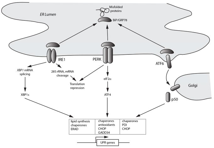
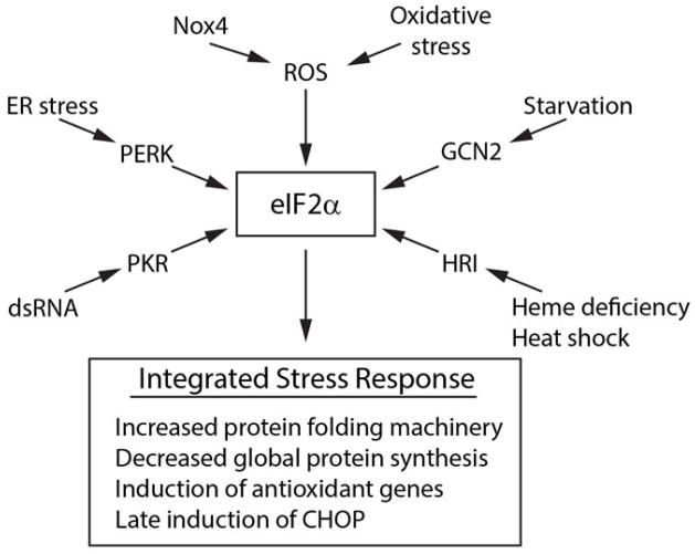
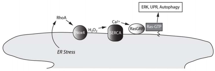
The endoplasmic reticulum (ER) produces the vast majority of all proteins secreted into the extracellular space, including hormones and cytokines, as well as cell surface receptors and other proteins which interact with the environment. Accordingly, this organelle controls essentially all vital links to a cell's external milieu, responding to systemic metabolic, inflammatory, endocrine, and mechanical stimuli. The central role the ER plays in meeting protein synthetic and quality control requirements in the face of such demands is matched by an extensive and versatile ER stress response signaling network. ROS mediate several critical aspects of this response. Nox4, an ER resident capable of producing ROS, acts as a proximal signaling intermediate to transduce ER stress-related conditions to the unfolded protein response, a homeostatic corrective mechanism. However, chronic ER stress caused by unrelenting internal or external demands produces a secondary rise in ROS, generally resulting in cell death. Sorting out the involvement of ROS at different levels of the ER stress response in specific cell types is key to understanding the molecular basis for chronic diseases such as atherosclerosis, hypertension, and diabetes. Here, we provide an overview of ER stress signaling with an emphasis on the role of ROS.

Keywords: Atherosclerosis; Autophagy; Hypertension; Nox4; Oxidative stress; Ras.


Figures

Figure 1
Figure 1. Overview of the unfolded protein response
Schematic shows titration of the chaperone BiP/Grp78 from the three main ER sensors IRE1 (inositol requiring enzyme 1), PERK (PKR-like eukaryotic initiating factor α kinase), and ATF6 (activating transcription factor 6) by misfolded proteins. The IRE1 pathway, through production of the alternatively translated product XBP1s, induces genes involved in lipid biosynthesis, ER-associated protein degradation (ERAD), and chaperones, to promote ER biogenesis, protein refolding, and disposal of damaged proteins. The PERK pathway, largely through induction of the transcription factor ATF4, additionally induces antioxidant-related genes and the phosphatase cofactor GADD34, which dephosphorylates eIF2α and deactivates the unfolded protein response (UPR). Release of ATF6 from BiP exposes Golgi localization signals, and ATF6 subsequently undergoes regulated intramembrane proteolysis to release a 50 kDa active transcription factor that targets genes involved in ER biogenesis and protein refolding.
Figure 2
Figure 2. The integrated stress response
Schematic shows the four main eIF2α kinases PERK, GCN2 (general control non-derepressible 2), PKR, and HRI (heme-regulated inhibitor) which respond to different stimuli to activate events downstream of eIF2α. eIF2α also responds to ROS either through Nox4 in response to ER stress, or to oxidative stress through undefined mechanisms.
Figure 3
Figure 3. Dual role of ROS in ER stress signaling
The left side of the panel depicts homeostatic signaling which involves ROS as signaling intermediates that report ER stress to the UPR, which in turn mitigates ER stress. Nox4 is shown as an important source of ROS in this capacity. In the event that ER stress is not relieved over time (depicted by the stop watch), delayed expression of proteins such as CHOP initiate a secondary rise in ROS (right side of the panel). This secondary increase in ROS appears to arise in part from induction of ER oxidase 1 (ERO1) and in part from calcium transfer across specialized ER-mitochondria contact sites with release of ROS, together contributing to cell death.
Figure 4
Figure 4. ROS in the stressed ER
a. Human umbilical vein endothelial cell expressing the H2O2 sensor HyPer targeted to the ER, after treatment with tunicamycin for 16 h. This photomicrograph shows a ratiometric image of HyPer demonstrating H2O2 elevations within swollen, dysmorphic ER vesicles with focal H2O2 accumulations particularly at the periphery of ER endomembranes. b. Nox4-GFP in live unfixed cell localizes to the reticular ER. Scale bars are 20 μm.
Figure 5
Figure 5. Nox4 signals ER stress
Schematic showing the role of Nox4 in relaying a local ER stress signaling pathway downstream of RhoA. Nox4-derived H2O2 oxidizes sulfhydryls of the sarcoendoplasmic reticulum calcium ATPase (SERCA), leading to an increase in cytosolic calcium, activation of the calcium sensitive ER guanyl exchange factor RasGRF, and GTP loading of Ras on-site on the ER surface. ER-localized Ras leads to homeostatic correction through the UPR, ERK activation, and autophagy.

References

    1. Abdulkarim B, Hernangomez M, Igoillo-Esteve M, Cunha DA, Marselli L, Marchetti P, Ladriere L, Cnop M. Guanabenz Sensitizes Pancreatic beta Cells to Lipotoxic Endoplasmic Reticulum Stress and Apoptosis. Endocrinology. 2017;158(6):1659–1670. - PubMed
    1. Anilkumar N, San Jose G, Sawyer I, Santos CX, Sand C, Brewer AC, Warren D, Shah AM. A 28-kDa splice variant of NADPH oxidase-4 is nuclear-localized and involved in redox signaling in vascular cells. Arterioscler Thromb Vasc Biol. 2013;33(4):e104–112. - PubMed
    1. Axe EL, Walker SA, Manifava M, Chandra P, Roderick HL, Habermann A, Griffiths G, Ktistakis NT. Autophagosome formation from membrane compartments enriched in phosphatidylinositol 3-phosphate and dynamically connected to the endoplasmic reticulum. J Cell Biol. 2008;182(4):685–701. - PMC - PubMed
    1. Back SH, Scheuner D, Han J, Song B, Ribick M, Wang J, Gildersleeve RD, Pennathur S, Kaufman RJ. Translation attenuation through eIF2alpha phosphorylation prevents oxidative stress and maintains the differentiated state in beta cells. Cell Metab. 2009;10(1):13–26. - PMC - PubMed
    1. Belousov VV, Fradkov AF, Lukyanov KA, Staroverov DB, Shakhbazov KS, Terskikh AV, Lukyanov S. Genetically encoded fluorescent indicator for intracellular hydrogen peroxide. Nat Methods. 2006;3(4):281–286. - PubMed

Publication types