. 2018 Sep;138(9):1925-1934.
doi: 10.1016/j.jid.2018.02.041. Epub 2018 Mar 17.

Epidermal FABP Prevents Chemical-Induced Skin Tumorigenesis by Regulation of TPA-Induced IFN/p53/SOX2 Pathway in Keratinocytes

Affiliations

Epidermal FABP Prevents Chemical-Induced Skin Tumorigenesis by Regulation of TPA-Induced IFN/p53/SOX2 Pathway in Keratinocytes

Yuwen Zhang et al. J Invest Dermatol. 2018 Sep.

Abstract

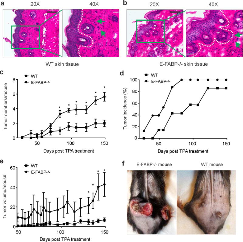
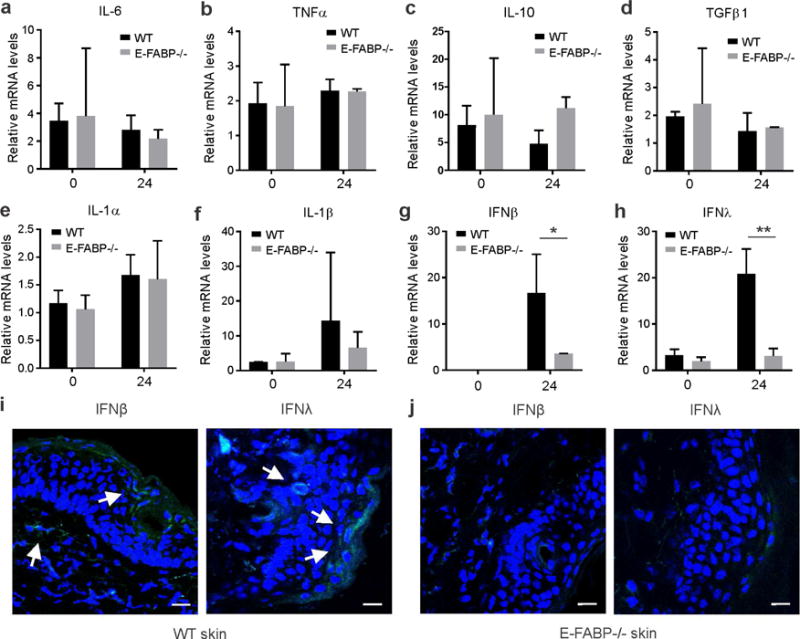
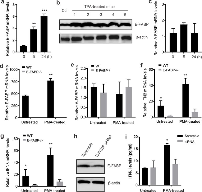
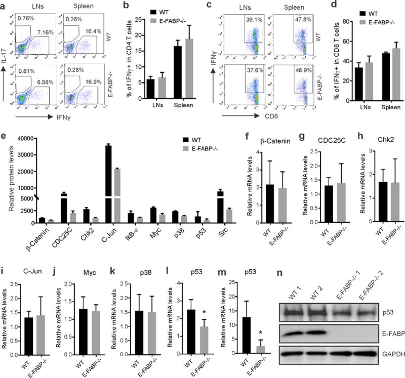
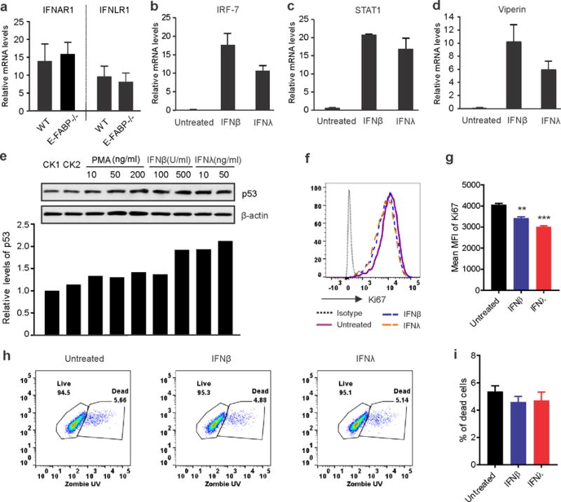
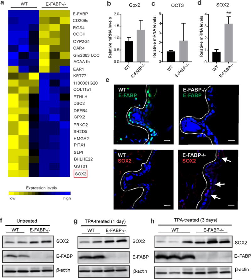
Skin lipids (e.g., fatty acids) are essential for normal skin functions. Epidermal FABP (E-FABP) is the predominant FABP expressed in skin epidermis. However, the role of E-FABP in skin homeostasis and pathology remains largely unknown. Herein, we utilized the 7,12-dimethylbenz(a)anthracene and 12-O-tetradecanolyphorbol-13-acetate-induced skin tumorigenesis model to assess the role of E-FABP in chemical-induced skin tumorigenesis. Compared to their wild-type littermates, mice deficient in E-FABP, but not adipose FABP, developed more skin tumors with higher incidence. 12-O-tetradecanolyphorbol-13-acetate functioning as a tumor promoter induced E-FABP expression and initiated extensive flaring inflammation in skin. Interestingly, 12-O-tetradecanolyphorbol-13-acetate -induced production of IFN-β and IFN-λ in the skin tissue was dependent on E-FABP expression. Further protein and gene expression arrays demonstrated that E-FABP was critical in enhancing IFN-induced p53 responses and in suppressing SOX2 expression in keratinocytes. Thus, E-FABP expression in skin suppresses chemical-induced skin tumorigenesis through regulation of IFN/p53/SOX2 pathway. Collectively, our data suggest an unknown function of E-FABP in prevention of skin tumor development, and offer E-FABP as a therapeutic target for improving skin innate immunity in chemical-induced skin tumor prevention.


Conflict of interest statement

Disclosure of Potential Conflicts of Interest

The authors state no conflict of interest.

Figures

Figure 1
Figure 1. Expression of E-FABP suppresses DMBA/TPA-induced skin tumor development
(a,b) Histological analysis (H&E staining) of keratinocyte proliferation and inflammatory cell infiltration in WT (a) and E-FABP−/− mice (b) treated with TPA for 7 days (infiltrated inflammatory cells pointed by green arrows) (scale bar = 100μM). c, Average skin tumor numbers per mouse in DMBA/TPA-treated WT and E-FABP−/− mice (n=12/group). d, The incidence of skin tumor formation in WT and E-FABP−/− mice treated with DMBA/TPA. e, Average tumor volume per mouse (mm3) in DMBA/TPA-treated WT and E-FABP−/− mice. f, Representative images of DMBA/TPA-induced tumors in WT and E-FABP−/− mice.
Figure 2
Figure 2. TPA treatment induces E-FABP- independent and dependent skin inflammation
Dorsal skin of WT and E-FABP mice was shaved and treated with or without 17 nmol TPA for 24 hours before collection for analyses of different types of cytokines and chemokines using quantitative PCR. a–h, relative expression of IL-6 (a), TNFα (b), IL-10(c), TGF1β (d), IL-1α (e), IL-1β (f), IFNβ (g) and IFNλ (h) was shown in untreated and treated WT and E-FABP−/− mice (n=4/group). Data are representative of three experiments. *, p<0.05 and **, p<0.01 as compared between WT and E-FABP−/− mice. (i,j) Analysis of expression of IFNβ and IFNλ in skin tissue of WT (i) and E-FABP−/− mice (j) treated with TPA for 7 days by confocal microscopy (IFNs: green; nucleus: blue; IFN producing cells: white arrows, scale bar = 10μM).
Figure 3
Figure 3. TPA treatment of skin upregulates E-FABP expression and E-FABP-dependent production of IFNβ and IFNλ
(a,b) Analyses of E-FABP expression in skin treated with/without TPA at the indicated time points by qPCR (a) or 3 days by Western blotting (b). c, Real-time PCR analysis of A-FABP mRNA expression in skin treated with or without TPA for different time points. d–g, Analysis of the expression of E-FABP (d), A-FABP (e), IFNβ (f) and IFNλ (g) in primary skin keratinocytes treated with or without PMA for 24 hours by quantitative real-time PCR (n=6/group). h, Western blotting analysis of E-FABP expression in MPEK keratinocytes transfected with scramble or E-FABP siRNA. i, Measurement of IFNλ levels in the supernatants of siRNA-transfected MPEK keratinocytes treated with/without PMA for 6 hours. Data are representative of three experiments (*, p<0.05; **, p<0.01).
Figure 4
Figure 4. E-FABP deficiency is associated with reduced p53 expression in keratinocytes
a–d, Draining lymph nodes (LNs) and spleen were collected from DMBA/TPA-treated tumor bearing mice and analyzed for IFNγ and IL-17 production by gating on CD4+ T cells (a) and IFNγ production in CD8+ T cells (c) by flow intracellular staining. Average percentage of IFNγ positive cells in CD4+ and CD8+ cells was shown in b and d, respectively (n=4/group). e, Cancer signaling proteins with relative fold > 2 between WT and E-FABP−/− skin tumors were shown by protein array analysis. f–l, qPCR analysis of relative expression of β-catenin (f), CDC25c (g), Chk2 (h), C-Jun (i), Myc (j), p38 (k) and p53 (l), in skin of naïve WT and E-FABP−/− mice. m, qPCR analysis of relative p53 levels in primary keratinocytes from WT and E-FABP−/− mice after PMA treatment for 24 hours (n=3/group, *p<0.05). n, Western blotting analysis of p53 expression in skin cell lysates after anti-p53 immunoprecipitation. The supernatants after p53-immunoprecipitation were directly analyzed for the expression of E-FABP and GAPDH.
Figure 5
Figure 5. IFNβ and IFNλ induce p53 expression in keratinocytes and inhibit their proliferation
a, qPCR analysis of IFNAR1 and IFNLR1 receptor expression in skin of naïve WT and E-FABP−/− mice. b–d, Analysis of expression of IRF-7 (b), STAT1 (c) and Viperin (d) in MPEK keratinocytes treated with IFNβ (500U/ml) or IFNλ (50ng/ml) for 8 hours. e, Western blotting analysis of p53 expression in MPEK keratinocytes stimulated with indicated concentrations of PMA, IFNβ and IFNλ. Relative protein levels are shown in the low panel. f–i, Flow cytometric analyses of Ki67 expression (f) and cell death (h) of MPEK keratinocytes treated with/without IFNβ or IFNλ. Mean fluorescent intensity (MFI) and average percentage of dead cells are shown in panel g and i, respectively. Results are representative of three times of experiments. (**, p<0.01, ***, p<0.001).
Figure 6
Figure 6. SOX2 expression is upregulated in skin of E-FABP deficiency mice
a, Heatmap of differentially expressed genes with fold >3 in tumors from WT and E-FABP−/−mice. b–d, Real-time PCR analysis of expression of Gpx2 (b), OCT3 (c) and SOX2 (d) in primary keratinocytes from WT and E-FABP−/− mice (n=3/group, **, p<0.01 as compared to WT mice). e, Confocal microscopic analysis of expression of E-FABP (green color) and SOX2 (red color) in keratinocytes of skin epidermis from WT and E-FABP deficiency mice (scale bar = 10μM). f–h, Western blotting analysis of SOX2 and E-FABP expression in skin of untreated naïve mice (f), mice treated with TPA for 1 day (g) and mice treated with TPA for 3 days (h).

References

    1. Abel EL, Angel JM, Kiguchi K, DiGiovanni J. Multi-stage chemical carcinogenesis in mouse skin: fundamentals and applications. Nat Protoc. 2009;4:1350–1362. - PMC - PubMed
    1. Boumahdi S, Driessens G, Lapouge G, Rorive S, Nassar D, Le MM, et al. SOX2 controls tumour initiation and cancer stem-cell functions in squamous-cell carcinoma. Nature. 2014;511:246–250. - PubMed
    1. Brosh R, Assia-Alroy Y, Molchadsky A, Bornstein C, Dekel E, Madar S, et al. p53 counteracts reprogramming by inhibiting mesenchymal-to-epithelial transition. Cell Death Differ. 2013;20:312–320. - PMC - PubMed
    1. Chen HM, Tanaka N, Mitani Y, Oda E, Nozawa H, Chen JZ, et al. Critical role for constitutive type I interferon signaling in the prevention of cellular transformation. Cancer Sci. 2009;100:449–456. - PMC - PubMed
    1. Crochemore C, Michaelidis TM, Fischer D, Loeffler JP, Almeida OF. Enhancement of p53 activity and inhibition of neural cell proliferation by glucocorticoid receptor activation. FASEB J. 2002;16:761–770. - PubMed

Publication types

MeSH terms