. 2018 Jun 1;78(11):3075-3086.
doi: 10.1158/0008-5472.CAN-17-3024. Epub 2018 Mar 20.

Cotargeting BCL-2 and PI3K Induces BAX-Dependent Mitochondrial Apoptosis in AML Cells

Affiliations

Cotargeting BCL-2 and PI3K Induces BAX-Dependent Mitochondrial Apoptosis in AML Cells

Mohamed Rahmani et al. Cancer Res. .

Abstract

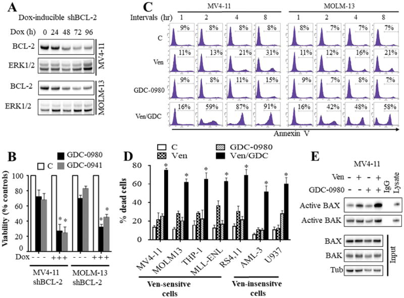
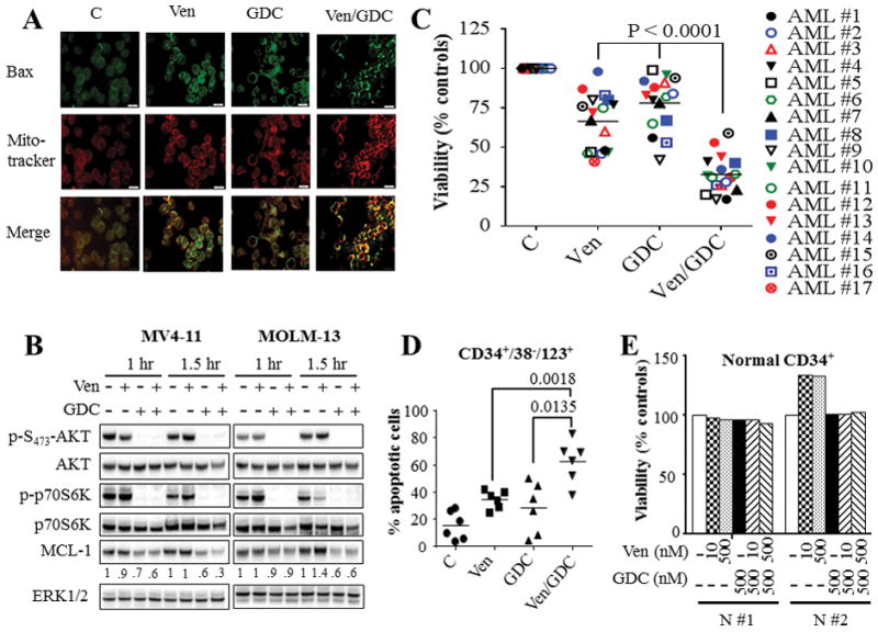
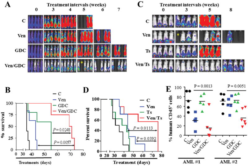
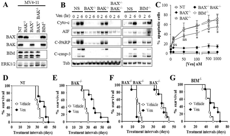
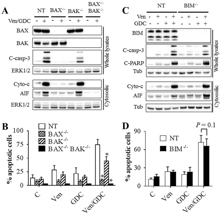
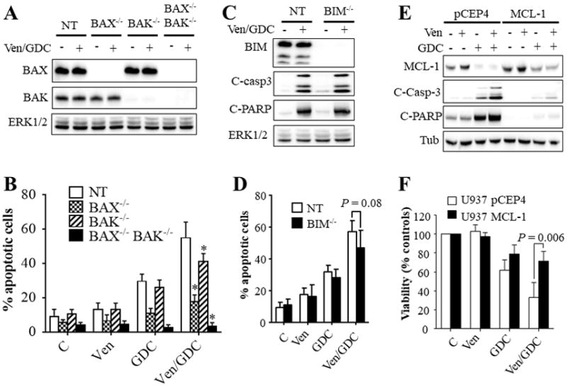
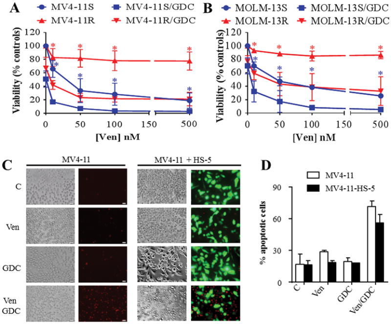
Inhibitors targeting BCL-2 apoptotic proteins have significant potential for the treatment of acute myeloid leukemia (AML); however, complete responses are observed in only 20% of patients, suggesting that targeting BCL-2 alone is insufficient to yield durable responses. Here, we assessed the efficacy of coadministration of the PI3K/mTOR inhibitor GDC-0980 or the p110β-sparing PI3K inhibitor taselisib with the selective BCL-2 antagonist venetoclax in AML cells. Tetracycline-inducible downregulation of BCL-2 significantly sensitized MV4-11 and MOLM-13 AML cells to PI3K inhibition. Venetoclax/GDC-0980 coadministration induced rapid and pronounced BAX mitochondrial translocation, cytochrome c release, and apoptosis in various AML cell lines in association with AKT/mTOR inactivation and MCL-1 downregulation; ectopic expression of MCL-1 significantly protected cells from this regimen. Combined treatment was also effective against primary AML blasts from 17 patients, including those bearing various genetic abnormalities. Venetoclax/GDC-0980 markedly induced apoptosis in primitive CD34+/38-/123+ AML cell populations but not in normal hematopoietic progenitor CD34+ cells. The regimen was also active against AML cells displaying intrinsic or acquired venetoclax resistance or tumor microenvironment-associated resistance. Either combinatorial treatment markedly reduced AML growth and prolonged survival in a systemic AML xenograft mouse model and diminished AML growth in two patient-derived xenograft models. Venetoclax/GDC-0980 activity was partially diminished in BAK-/- cells and failed to induce apoptosis in BAX-/- and BAX-/-BAK-/- cells, whereas BIM-/- cells were fully sensitive. Similar results were observed with venetoclax alone in in vitro and in vivo systemic xenograft models. Collectively, these studies demonstrate that venetoclax/GDC-0980 exhibits potent anti-AML activity primarily through BAX and, to a lesser extent, BAK. These findings argue that dual BCL-2 and PI3K inhibition warrants further evaluation in AML.Significance: Combined treatment with clinically relevant PI3K and BCL-2 inhibitors may prove effective in the treatment of acute myeloid leukemia. Cancer Res; 78(11); 3075-86. ©2018 AACR.


Conflict of interest statement

Conflict of interest disclosures: J.L, and D.S are employees of AbbVie and Genentech respectively. All other authors have no conflict of interest to disclose.

Figures

Figure 1
Figure 1. Dual inhibition of BCL-2 and PI3K pathway potently induced apoptosis in AML cells in association with a pronounced change in BAX conformation
A) western blot analysis in MV4-11 and MOLM-13 cells displaying inducible knock-down of BCL-2 following exposure to 1 μg/ml doxycycline (Dox). B) These cells were left untreated or treated with doxycycline for 48 hr, and then exposed to the PI3K inhibitors GDC-0980 (500 nM) or GDC-0941 (1 μM) for 24 hr after which cell growth and viability was assessed by a CellTiter-Glo luminescent assay. Error Bars: S.D of 3 independent experiments; *, P < 0.05 in each case for values obtained in the presence vs absence of doxycycline. C) MV4-11 and MOLM-13 cells were exposed to venetoclax (10 nM) and GDC-0980 (500 nM) alone or together for the designated intervals after which the extent of apoptosis was determined by Annexin V staining. D) Various AML cell lines were exposed to ABT-199 and/or GDC-0980 for 24 hr after which the extent of cell death was determined employing Annexin V/PI staining. The agent concentrations used were: ABT-199: 2.5 nM for RS4,11; 10 nM for MV4-11 and MOLM-13; 50 nM for MLL-ENL; 100 nM for THP-1; and 500 nM for U937 and OCI-AML3 cells. GDC-0980: 1000 nM for U937 and RS4,11 cells; 500 nM for all other cell lines. Error Bars, S.D for at least 3 independent experiments; P < 0.003 for combined treatment compared to either agent alone for each cells. E) MV4-11 cells were exposed to venetoclax and/or GDC-0980 for 2 hr after which BAX and BAK conformational changes were assessed (top panel). Western blot analysis was also performed on input lysates (bottom panel).
Figure 2
Figure 2. Venetoclax/GDC-0980 inactivates AKT/mTOR/p70S6K, down-regulates MCL-1, triggers BAX mitochondrial translocation, and effectively kills primary AML blasts including CD34+/38/123+ AML progenitor but not normal CD34+ cells
(A) MV4-11 cells were exposed to 10 nM venetoclax (Ven) and/or 500 nM GDC-0980 (GDC) for 2 hr after which BAX translocation to the mitochondria was assessed using IX71 Olympus microscope. (B) MV4-11 and MOLM-13 cells were treated with 10 nM venetoclax (Ven) and/or 500 nM GDC-0980 (GDC) for the indicated intervals, after which cells were lysed and the lysates were subjected to western blot analysis. Densitometry analysis was performed on MCL-1 blots using Image Studio lite Software (Li-Cor Biosciences), and values were normalized for ERK1/2 loading controls. (C) Assessment of cell viability using Annexin V/7-AAD or CellTiter-Glo luminescent assays for 17 primary AML specimens with a preponderance of blasts (≥ 80%) following 16 hr treatment with venetoclax and GDC-0980 alone or together. As in the case of cell lines, primary AML samples exhibited heterogeneous responses to venetoclax. Concentrations of venetoclax were selected based upon marginal toxicity when administered alone and clinical relevance. Venetoclax concentrations varied between 10 – 100 nM for patients #1–10, and between 200 – 2000 nM for patients #11 – 17. GDC-0980 was administered at concentrations varying between 200 nM and 1000 nM. The median values for combined treatment were significantly lower than values for either agent alone (P < 0.0001 in each case). (D) Primary AML blasts from 6 patients were treated as in (C) for 16 hr after which the extent of the cell death was assessed selectively in AML progenitor CD34+/CD38/CD123+ cells by Annexin V/7-AAD staining. The median values were significantly lower for combined treatment compared to either agent alone (P < 0.0018; P < 0.0135 for venetoclax and GDC-0980 respectively). (E) Normal hematopoietic mononuclear cells were isolated from umbilical cord blood of 2 subjects (N #1 and N #2) and were exposed to the designated concentrations of venetoclax and GDC-0980 alone or in combination for 16 hr after which viability was assessed selectively in the CD34+ cell population by Annexin V/7-AAD staining.
Figure 3
Figure 3. Co-treatment with venetoclax and GDC-0980 exhibits potent in vivo anti-AML activity
NOD/SCID-γ mice were inoculated with 3 × 10⁶ luciferase-expressing MV4-11 cells via tail vein injection. 1 week later, mice were treated with regimens consisting of oral administration of venetoclax, GDC-0980, or taselisib once every day, 6 days a week as follow. (A) Mice were treated with venetoclax (80 mg/kg), GDC-0980 (5 mg/kg for 2 weeks, then 10 mg/kg until day 55) alone or in combination, and AML progression was monitored by the IVIS 200 imaging system. (B) Survival analysis of mice using Kaplan-Meier survival plot. These studies involved 5–6 mice/condition; combined treatment significantly prolonged mouse survival compared to either agent alone (P = 0.0248, and P = 0.0057 for combination versus GDC-0980 and venetoclax respectively, log-rank test). (C) Mice (8 mice/condition) were treated with venetoclax (80 mg/kg) and/or taselisib (2.5 mg/kg), and subjected to IVIS 200 imaging. (D) Kaplan-Meier survival analysis. Venetoclax/taselisib significantly prolonged mouse survival compared to either agent alone (P = 0.0113, and P = 0.0392 for combination versus taselisib and venetoclax respectively, log-rank test). * indicate mouse accidental death; these mice were excluded from survival analysis. (E) NOD/SCID-γ mice were inoculated with primary AML blasts (5 × 10⁶ cells) isolated from 2 patients with AML. 2 weeks later, mice were treated with venetoclax (80 mg/kg) and/or GDC-0980 (5 mg/kg) po qd 6 days/week for 3 weeks. Mice were left untreated for additional 3 months after which they were sacrificed and the percentages of HCD45+ cells in the bone marrow were assessed. Each dot represents an individual mouse (n = 5 – 6 per group). The means of different groups are statistically different for both patients AML1; P = 0.0013; AML2; P = 0.0051; One-way ANOVA.
Figure 4
Figure 4. Functional roles of BAX, BAK, and BIM in venetoclax activity in vitro and in vivo
(A) western blot analysis in untreated MV4-11 cells in which BAX (BAX−/−), BAK (BAK−/−), double BAX/BAK (BAX−/−BAK−/−), or BIM (BIM−/−) were knocked out using CRISPR technology as described in Methods or non-targeting (NT) control cells. (B) These cell were exposed to 100 nM venetoclax (Ven) for 2 or 6 hr after which cytosolic fractions were isolated and subjected to western blot analysis. (C) Cells were treated with the designated concentrations of venetoclax for 24 hr after which the extent of apoptosis was determined using Annexin V staining. Error Bars, S.D for at least 3 independent experiments.*, P < 0.05; ns = non-significant for BAK−/− versus NT cells. Values obtained for BAX−/− or BAX−/−BAK−/− cells before versus after venetoclax treatment were not significantly different, P > 0.05 in each case. (D–G) NOD/SCID-γ mice were inoculated with 3 × 10⁶ BAX−/−, BAK−/−, BAX−/−BAK−/−, BIM−/−, or NT control MV4-11 cells via tail vein. One week later, mice were treated orally with 75 mg/kg venetoclax (Ven) once every day, 6 days a week. Mouse survival was analyzed by Kaplan-Meier survival plot. Survival of mice with Bax−/− or Bax−/−Bak−/−-derived xenografts are both included in Figure 4F. These studies involved 8 mice/condition. Venetoclax significantly prolonged mouse survival in mice with NT, BAK−/−, or BIM−/− cells, P = 0.0138, P = 0.0015, and P = 0.0117 respectively. In contrast, no survival benefit was observed in mice with BAX−/− or BAX−/−BAK−/− cells.
Figure 5
Figure 5. Role of BAX, BAK, and BIM in venetoclax/GDC-0980 lethality in venetoclax-sensitive MV4-11 cells
Cells in which BAX, BAK, double BAX/BAK (A–B), or BIM (C–D) were knocked out using CRISPR or non-targeting (NT) control cells were exposed to 10 nM venetoclax and/or 500 nM GDC-0980. Cells were either lysed after 2 hr and subjected to western blot analysis (A–C), or analyzed for the extent of apoptosis at 5 hr using Annexin V staining (B–D). Error Bars, S.D for at least 4 independent experiments. For B: P = 0.0004; P = 0.0027; P = 0.0002 for BAX−/−, BAK−/−, or BAX−/−BAK−/− cells respectively compared to NT cells; for D: P = 0.1307 for BIM−/− cells vs NT cells.
Figure 6
Figure 6. Role of BAX, BAK, and BIM in venetoclax/GDC-0980 lethality in venetoclax-insensitive U937 cells
Cells exhibiting knockout of BAX, BAK, double BAX/BAK (A–B), or BIM (C–D), or non-targeting (NT) control cells were exposed to venetoclax (500 nM) and/or GDC-0980 (1.5 μM) for 16 hr. The cells were then lysed, and the lysates subjected to western blot analysis (A, C). Alternatively, the extent of apoptosis was determined using the Annexin V staining (B, D). Error Bars, S.D of at least 3 independent experiments. For B: P = 0.0005; P = 0.0223; P < 0.0001 for BAX−/−, BAK−/−, or BAX−/−BAK−/− cells respectively compared to NT cells; for D: P = 0.08. (E–F) western blot analysis (E) and CellTiter-Glo luminescent viability assay (F) on U937 cells ectopically expressing MCL-1 or the empty vector pCEP4 following exposure to venetoclax (500 nM) and/or GDC-0980 (1.5 μM). For (F), Error Bars, S.D for 4 independent experiments; P = 0.006.
Figure 7
Figure 7. Venetoclax/GDC-0980 is effective in AML cells exhibiting various forms of venetoclax resistance
(A–B) venetoclax-sensitive (MV4-11S, MOLM-13S) or venetoclax-resistant (MV4-11R, MOLM-13R) cells were exposed to the designated concentrations of venetoclax alone or in combination with 500 nM GDC-0980 for 24 hr after which cell growth and viability was assessed using the CellTiter-Glo luminescent assay. Error Bars, S.D for at least 3 independent experiments. P < 0.01 for combined treatment compared to venetoclax alone for each concentration in each cell line. (C) MV4-11 cells were cultured in the presence or absence of GFP-labeled HS-5 stromal cells for 48 hr then exposed to venetoclax ± GDC-0980 (10 and 500 nM respectively) for 5 hr after which, cells were stained with 7-AAD for 30 min and photographed as described in Methods. Alternatively, the extent of apoptosis was determined in MV4-11 cells (GFP-negative population) using the Annexin V-APC staining (Fig. 7D). Error Bars, S.D for 4 independent experiments.

References

    1. Delbridge AR, Strasser A. The BCL-2 protein family, BH3-mimetics and cancer therapy. Cell Death Differ. 2015;22:1071–80. - PMC - PubMed
    1. Oltersdorf T, Elmore SW, Shoemaker AR, Armstrong RC, Augeri DJ, Belli BA, et al. An inhibitor of Bcl-2 family proteins induces regression of solid tumours. Nature. 2005;435:677–81. - PubMed
    1. Park C-M, Bruncko M, Adickes J, Bauch J, Ding H, Kunzer A, et al. Discovery of an orally bioavailable small molecule inhibitor of prosurvival B-cell lymphoma 2 proteins. J Med Chem. 2008;51:6902–15. - PubMed
    1. Wilson WH, O’Connor OA, Czuczman MS, LaCasce AS, Gerecitano JF, Leonard JP, et al. Navitoclax, a targeted high-affinity inhibitor of BCL-2, in lymphoid malignancies: a phase 1 dose-escalation study of safety, pharmacokinetics, pharmacodynamics, and antitumour activity. Lancet Oncol. 2010;11:1149–59. - PMC - PubMed
    1. Gandhi L, Camidge DR, Ribeiro de Oliveira M, Bonomi P, Gandara D, Khaira D, et al. Phase I study of Navitoclax (ABT-263), a novel Bcl-2 family inhibitor, in patients with small-cell lung cancer and other solid tumors. J Clin Oncol. 2011;29:909–16. - PMC - PubMed

Publication types

MeSH terms

Substances