. 2018 Apr;67(4):537-547.
doi: 10.2337/dbi17-0040.

New Understanding of β-Cell Heterogeneity and In Situ Islet Function

Affiliations

New Understanding of β-Cell Heterogeneity and In Situ Islet Function

Richard K P Benninger et al. Diabetes. 2018 Apr.

Abstract

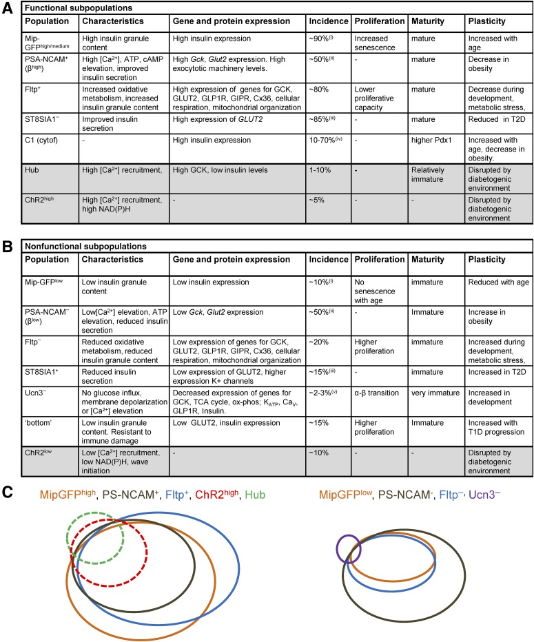
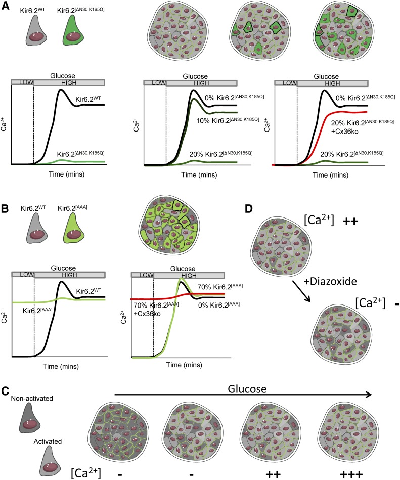
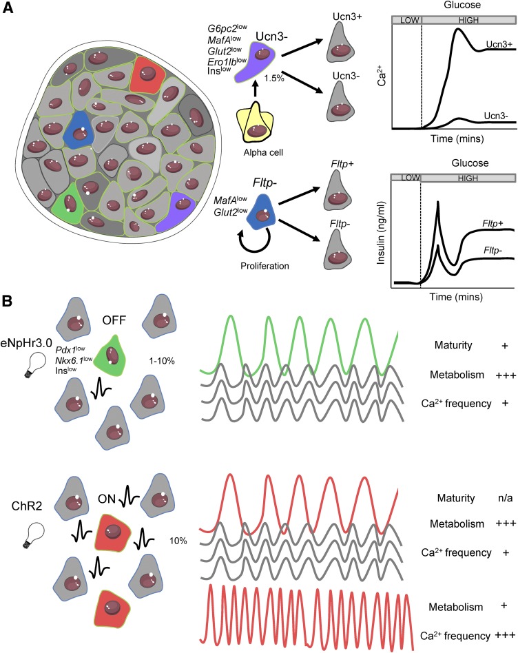
Insulin-secreting β-cells are heterogeneous in their regulation of hormone release. While long known, recent technological advances and new markers have allowed the identification of novel subpopulations, improving our understanding of the molecular basis for heterogeneity. This includes specific subpopulations with distinct functional characteristics, developmental programs, abilities to proliferate in response to metabolic or developmental cues, and resistance to immune-mediated damage. Importantly, these subpopulations change in disease or aging, including in human disease. Although discovering new β-cell subpopulations has substantially advanced our understanding of islet biology, a point of caution is that these characteristics have often necessarily been identified in single β-cells dissociated from the islet. β-Cells in the islet show extensive communication with each other via gap junctions and with other cell types via diffusible chemical messengers. As such, how these different subpopulations contribute to in situ islet function, including during plasticity, is not well understood. We will discuss recent findings revealing functional β-cell subpopulations in the intact islet, the underlying basis for these identified subpopulations, and how these subpopulations may influence in situ islet function. Furthermore, we will discuss the outlook for emerging technologies to gain further insight into the role of subpopulations in in situ islet function.


Figures

Figure 1
Figure 1
Summary of previously identified β-cell subpopulations using biomarker or single-cell analyses. A: Summary of functionally competent (functional) cell subpopulations, including their functional and gene/protein expression profile, incidence, and how they change in conditions associated with diabetes. B: Summary of less functional (nonfunctional) cell subpopulations. For A and B, shaded rows are functionally defined via optogenetics in situ. C: Schematic suggesting qualitative overlap between several identified subpopulations. Dashed lines indicate those defined via optogenetics in situ. Note the strong overlap between most identified larger populations, but the low-incidence functional hub cell subpopulation and nonfunctional Ucn3 subpopulation show relatively lower overlap. This may reflect the number and nature of the markers measured, as well as the functional readout. ox-phos, oxidative phosphorylation; T1D, type 1 diabetes; TCA, tricarboxylic acid. (i) GFPhigh increases from 10–40% with age (from 5–9 weeks to 16–40 weeks) and GFPlow decreases from 50 to 10% with age. (ii) PSA-NCAM+ is defined as top 50% expression. (iii) Incidence was 50% in donors with type 2 diabetes (T2D). (iv) Incidence was <30% in donors with T2D. (v) Ucn3, insulin+ is located at the islet periphery. Adapted from Servier Medical Art under a CC-BY3.0 license (https://creativecommons.org/licenses/by/3.0/).
Figure 2
Figure 2
Example for how heterogeneous cells can interact. A: Introducing Kir6.2[ΔN30,K185Q] into β-cells renders them unresponsive to glucose. With a small (∼10%) population of these less functional cells in the islet (dark green), glucose responsiveness is unchanged. However, with ∼20% of these cells, glucose responsiveness is lost. Glucose responsiveness is recovered by electrically isolating (Cx36ko) these less functional cells. B: Conversely, if a large proportion (∼70%) of hyperexcitable Kir6.2[AAA] cells are introduced into the islet (light green), glucose responsiveness remains, although this is lost when electrical coupling is lost (Cx36ko). C: In the native islet, the number of intrinsically activated (light gray) cells increases as glucose concentration rises. However, only when a certain threshold of cells are activated does Ca2+ elevate as a result of insufficient numbers of nonactivated cells (dark gray), analogous to A and B. D: The presence of activated and nonactivated cells can be further revealed by the KATP opener diazoxide, which renders only a small number of cells intrinsically inactive as observed in the absence of GJ coupling but fully abolishes Ca2+ elevations in the presence of GJ coupling. Adapted from Servier Medical Art under a CC-BY3.0 license (https://creativecommons.org/licenses/by/3.0/).
Figure 3
Figure 3
Heterogeneity in the intact islet. A: Different β-cell subpopulations coexist within the islet, each characterized by different gene/protein expression patterns, morphological markers, glucose responsiveness, insulin secretion, and proliferative capacity and function (Fig. 1). Fltp proliferative (blue) and Ucn3 transdifferentiating (purple) less functional immature populations are likely nonelectrically coupled and thus do not significantly affect islet-wide responses to glucose. B: Optogenetic mapping reveals highly functional β-cell subpopulations with varying identity markers, metabolic properties, and Ca2+ responses, but with the ability to exert disproportionate control over coordination and intraislet Ca2+ responses. This includes eNpHR3.0-silenced β-cells (hub cells, green) in which halorhodopsin activation and membrane hyperpolarization disproportionally silence the islet and ChR2-activated cells (red) in which ChR2 activation and membrane depolarization disproportionally activate the islet. A population of cells in which ChR2 activation has little effect also shows pacemaker-like characteristics owing to their higher intrinsic oscillation frequency. Adapted from Servier Medical Art under a CC-BY3.0 license (https://creativecommons.org/licenses/by/3.0/).

Similar articles

Cited by

References

    1. Pipeleers DG. Heterogeneity in pancreatic beta-cell population. Diabetes 1992;41:777–781 - PubMed
    1. Salomon D, Meda P. Heterogeneity and contact-dependent regulation of hormone secretion by individual B cells. Exp Cell Res 1986;162:507–520 - PubMed
    1. Misler S, Falke LC, Gillis K, McDaniel ML. A metabolite-regulated potassium channel in rat pancreatic B cells. Proc Natl Acad Sci U S A 1986;83:7119–7123 - PMC - PubMed
    1. Kiekens R, In ’t Veld P, Mahler T, Schuit F, Van De Winkel M, Pipeleers D. Differences in glucose recognition by individual rat pancreatic B cells are associated with intercellular differences in glucose-induced biosynthetic activity. J Clin Invest 1992;89:117–125 - PMC - PubMed
    1. Piston DW, Knobel SM, Postic C, Shelton KD, Magnuson MA. Adenovirus-mediated knockout of a conditional glucokinase gene in isolated pancreatic islets reveals an essential role for proximal metabolic coupling events in glucose-stimulated insulin secretion. J Biol Chem 1999;274:1000–1004 - PubMed

Publication types