. 2018 Apr 3;115(14):E3173-E3181.
doi: 10.1073/pnas.1713510115. Epub 2018 Mar 20.

PDGFRα+ pericryptal stromal cells are the critical source of Wnts and RSPO3 for murine intestinal stem cells in vivo

Affiliations

PDGFRα+ pericryptal stromal cells are the critical source of Wnts and RSPO3 for murine intestinal stem cells in vivo

Gediminas Greicius et al. Proc Natl Acad Sci U S A. .

Abstract

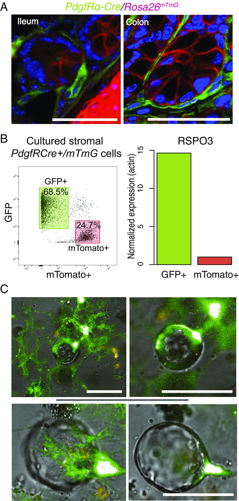
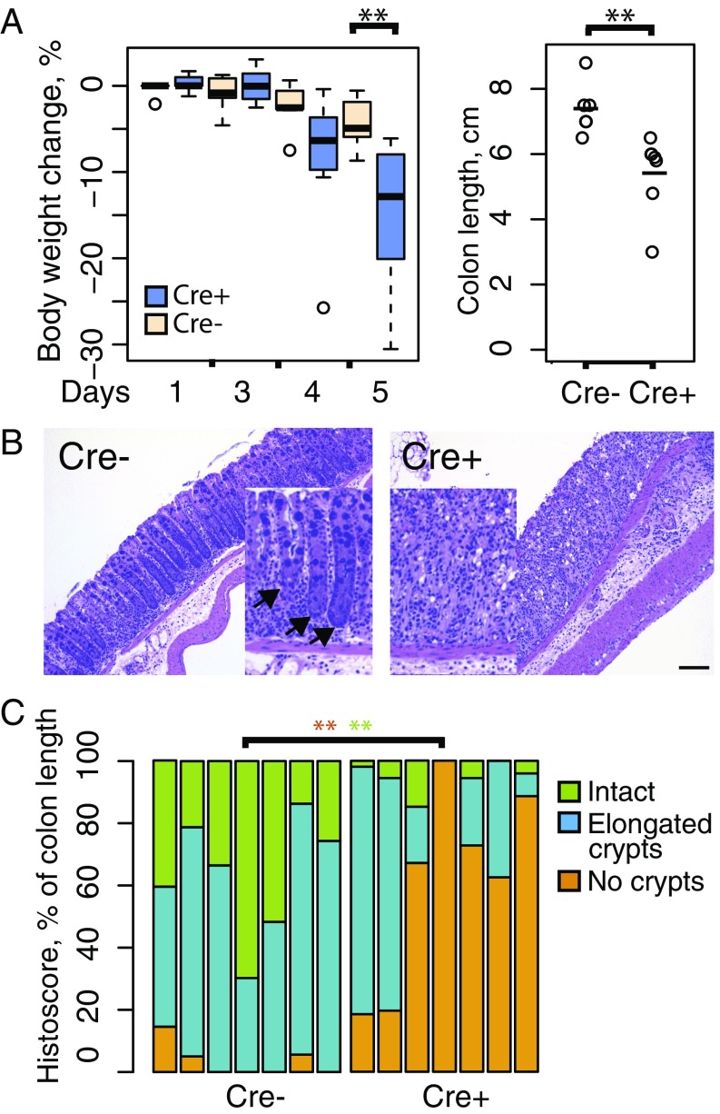
Wnts and R-spondins (RSPOs) support intestinal homeostasis by regulating crypt cell proliferation and differentiation. Ex vivo, Wnts secreted by Paneth cells in organoids can regulate the proliferation and differentiation of Lgr5-expressing intestinal stem cells. However, in vivo, Paneth cell and indeed all epithelial Wnt production is completely dispensable, and the cellular source of Wnts and RSPOs that maintain the intestinal stem-cell niche is not known. Here we investigated both the source and the functional role of stromal Wnts and RSPO3 in regulation of intestinal homeostasis. RSPO3 is highly expressed in pericryptal myofibroblasts in the lamina propria and is several orders of magnitude more potent than RSPO1 in stimulating both Wnt/β-catenin signaling and organoid growth. Stromal Rspo3 ablation ex vivo resulted in markedly decreased organoid growth that was rescued by exogenous RSPO3 protein. Pdgf receptor alpha (PdgfRα) is known to be expressed in pericryptal myofibroblasts. We therefore evaluated if PdgfRα identified the key stromal niche cells. In vivo, Porcn excision in PdgfRα+ cells blocked intestinal crypt formation, demonstrating that Wnt production in the stroma is both necessary and sufficient to support the intestinal stem-cell niche. Mice with Rspo3 excision in the PdgfRα+ cells had decreased intestinal crypt Wnt/β-catenin signaling and Paneth cell differentiation and were hypersensitive when stressed with dextran sodium sulfate. The data support a model of the intestinal stem-cell niche regulated by both Wnts and RSPO3 supplied predominantly by stromal pericryptal myofibroblasts marked by PdgfRα.

Keywords: R-spondins; Wnt signaling; intestine; myofibroblasts; stroma.


Conflict of interest statement

The authors declare no conflict of interest.

Figures

Fig. 1.
Fig. 1.
RSPO3 is more potent than RSPO1 in activating a β-catenin reporter and supporting intestinal organoid cultures. (A) STF3A cells were transfected with the indicated amounts of RSPO1 and RSPO3 expression plasmids, and the resultant Wnt/β-catenin–dependent luciferase signal was measured as described. (B) Comparative RSPO1-Myc and RSPO3-Myc expression levels in transfected STF3A cells assessed by anti-MYC immunoblot. Mw, molecular weight. (C) Recombinant RSPO3 and RSPO1 were added at the indicated concentrations to HEK293 STF3A cells with robust endogenous Wnt signaling as described. Data from two independent experiments with similar results were normalized and merged, with the activity induced by 200 ng/mL RSPO1 set as 100%. (D) RSPO3 is more potent than RSPO1 in promoting organoid growth. Organoids were cultured with the indicated concentration of RSPO and counted in three wells per group on day 5. Data from two independent experiments were equalized using the organoid count in the RSPO1 500 ng/mL group as 100% response. (E) RSPO1- and RSPO3-stimulated organoids have similar morphologic features. Organoids were cultured in the presence of the indicated R-spondin concentrations and photographed on day 5. (Scale bar: 100 μm.) (F) RSPO3 is more potent than RSPO1 in regulating differentiation in intestinal epithelial organoids. The expression of the indicated differentiation markers from the organoids in D was assessed at day 5, normalized to β-actin expression levels. The figures combine two independent experiments, equalized by setting the expression in the RSPO3 100 ng/mL group as 100% response. *P < 0.05, Wilcoxon rank sum test.
Fig. 2.
Fig. 2.
The majority of RSPO3-expressing cells are pericryptal myofibroblasts. (A) Frozen sections of normal mouse colon or small intestine as indicated were labeled with antibodies specific for RSPO3 (red) and E-cadherin (green) or with nonspecific primary IgGs from the respective species; nuclei were counterstained with Hoechst. Pericryptal cells positive for RSPO3 are indicated with white arrows. (Scale bars: 50 μm.) (B, Upper) RSPO3 is coexpressed with SMA. Purified stromal cells were cultured for 7 d, fixed, and costained with antibodies specific for RSPO3 (yellow) and the myofibroblast-specific marker SMA (red), as well as desmin (red) and fibroblast-specific protein 1 (FSP1) (red), as indicated. (Scale bars: 90 μm.) (Lower) Quantitation of costaining using Columbus software. At least 450 cells were quantified per group. The percentage of cells in each quadrant is indicated. The quantification was repeated three times with similar results.
Fig. 3.
Fig. 3.
Stromal RSPO3 and Wnts are critical for adult intestinal homeostasis. (A) Intestinal stromal cells contact Lgr5-expressing cells in vitro. mTomato-expressing stromal cells from RosamTmG mice were combined with purified epithelial crypts from Lgr5-GFP mice. The mixed cells were cultured without added RSPO for 5 d and then were imaged using an inverted Zeiss LSM 710 microscope. Intestinal stroma, Lgr5+ cells, and nuclei are labeled red, green, and blue, respectively. (Scale bar: 20 μm.) (B) Stromal RSPO3 is required for organoid growth. PORCN-deficient intestinal epithelial cells requiring Wnts from stroma were cultured either alone or in combination with intestinal stromal cells. Rspo3 expression was targeted using siRNA (Upper) or adenoviral-expressed Cre (Lower). Mock targeting was performed with scrambled siRNA or GFP-expressing adenovirus, respectively. In the rescue, RSPO3 (50 ng/mL) was added. Combined data from four siRNA and three viral-targeting experiments are presented. **P < 0.001, Wilcoxon rank sum test.
Fig. 4.
Fig. 4.
PdgfRα-Cre–driven GFP reporter activation labels pericryptal myofibroblasts. (A) Samples from ileum and colon of PdgfRα-Cre+/RosamTmG mice were fixed, prepared, and stained with GFP-specific antibodies as described in Methods. Nuclei were stained with DAPI. The image is representative of the intestinal crypts of the three mice analyzed. (Scale bars: 50 μm.) (B) PdgfRα-Cre–labeled cells express RSPO3. PdgfRα-Cre+/RosamTmG myofibroblasts were prepared, cultured for 10 d, and FACS-sorted into GFP+ and mTomato+ populations. (Left) The proportion of positive cells in the population is presented as a percentage in a respective gate. (Right) Rspo3 expression levels were analyzed by qRT-PCR and normalized to Actb. (C) PdgfRα-Cre–labeled cells support Porcn-deficient organoids. PdgfRα-Cre+/RosamTmG myofibroblasts were prepared, cultured for 10 d, and mixed with Porcn-deficient epithelial cells. The mixed cell cultures were incubated for 4 d and photographed thereafter. Images of two organoids at different focal planes are representative of two experiments with similar results. (Scale bars: 100 μm.)
Fig. 5.
Fig. 5.
Porcn ablation in PdgfRα-expressing cells inhibits crypt formation in the neonatal gut. Intestines from PdgfRα-Cre+/Porcnfl/fl mice were harvested on postnatal day 5 and processed as described in Methods. (A) Intestinal (Upper) and abdominal skin (Lower) tissues were subjected to H&E or EdU staining, respectively. The figure represents one pair of littermates representative of six littermates analyzed. Arrows indicate newly formed crypts. (Scale bars: 100 μm.) (B) Porcn excision in PdgfRα-Cre–lineage cells leads to decreased Axin2 expression levels (Left) and decreased crypt counts (Right). Crypts were counted using histological sections from five littermates in each group. (C) Porcn excision in PdgfRα-Cre–expressing cells leads to significantly decreased numbers of proliferating cells in the epithelial layer of lamina propria and stroma. Proliferating cells were counted in either the top layer of mucosa, which typically represents epithelia, or deeper stromal layers in samples from five littermates and are presented as a ratio to total Hoechst 33342-labeled nuclei. (D) Porcn excision in PdgfRα-Cre mice does not inhibit proliferation in bulbs of skin hair follicles. Images are representative of samples of abdominal skin of three littermates per group that were analyzed. (Scale bar: 100 μm.) *P < 0.05, **P < 0.01, Wilcoxon rank sum test.
Fig. 6.
Fig. 6.
RSPO3 excision in intestinal myofibroblasts does not perturb normal intestinal homeostasis. (A) PdgfRα-Cre+/Rspo3fl/fl mice express significantly less Rspo3. Samples from ileum (Left) and colon (Right) of male (turquoise) and female (orange) PdgfRα-Cre+/Rspo3fl/fl and PdgfRα-Cre/Rspo3fl/fl mice were collected and analyzed for RNA expression. All samples were normalized to HPRT and then normalized. Mean Ct values of 28.4–30.4, 30.6–31.6, and 26.9–27.4- were obtained for Rspo1, Rspo2, and Rspo3, respectively, in the Rspo3 nonexcised group. (B) PdgfRα-Cre+/Rspo3fl/fl mice have a decrease in lysozyme-producing Paneth cells but no decrease in intestinal crypt proliferation. Samples from EdU-pulsed PdgfRα-Cre+/Rspo3fl/fl and PdgfRα-Cre/Rspo3fl/fl mice were prepared and stained with EdU- and lysozyme-specific reagents. Pictures are representative of two mice analyzed from each group. (Scale bars: 100 μm.) (C) Rspo3 excision in PdgfRα-Cre–lineage cells leads to decreased abundance of Axin2, Lgr5, and lysozyme (Lys) mRNA in full-thickness slices. Expression levels were normalized to Hprt. *P < 0.05, **P < 0.01, Wilcoxon rank sum test.
Fig. 7.
Fig. 7.
Rspo3 excision in intestinal myofibroblasts predisposes to DSS-induced colitis. (A, Left) PdgfRα-Cre+/Rspo3fl/fl mice treated with DSS for 5 d rapidly lose weight. **P = 0.007, Wilcoxon rank sum test. (Right) Colon length was significantly shorter in knockout mice at the end of 5 d treatment. **P = 0.002, Wilcoxon rank sum test. (B) PdgfRα-Cre+/Rspo3 fl/fl mice display extensive inflammatory infiltrates in the lamina propria and inflammation-associated crypt loss after 5 d of DSS treatment (Right) compared with nontargeted controls (Left). (C) Quantification of inflammation-affected regions after 5 d of DSS treatment. Rspo3-deleted (labeled as Cre+) intestines indicate significantly higher crypt loss compared with Rspo3fl/fl or PdgfRα-Cre+/Rspo3wt controls (labeled as Cre−). **P = 0.0019 and P = 0.0032 for areas with no crypts and those with intact crypt morphology, respectively, Wilcoxon rank sum test.

References

    1. Clevers H. The intestinal crypt, a prototype stem cell compartment. Cell. 2013;154:274–284. - PubMed
    1. Kuhnert F, et al. Essential requirement for Wnt signaling in proliferation of adult small intestine and colon revealed by adenoviral expression of Dickkopf-1. Proc Natl Acad Sci USA. 2004;101:266–271. - PMC - PubMed
    1. Korinek V, et al. Depletion of epithelial stem-cell compartments in the small intestine of mice lacking Tcf-4. Nat Genet. 1998;19:379–383. - PubMed
    1. Yan KS, et al. Non-equivalence of Wnt and R-spondin ligands during Lgr5+intestinal stem-cell self-renewal. Nature. 2017;545:238–242. - PMC - PubMed
    1. Storm EE, et al. Targeting PTPRK-RSPO3 colon tumours promotes differentiation and loss of stem-cell function. Nature. 2016;529:97–100. - PubMed

Publication types

MeSH terms