. 2018 May 4;293(18):6762-6775.
doi: 10.1074/jbc.RA117.001163. Epub 2018 Mar 20.

The balance of interleukin (IL)-6, IL-6·soluble IL-6 receptor (sIL-6R), and IL-6·sIL-6R·sgp130 complexes allows simultaneous classic and trans-signaling

Affiliations

The balance of interleukin (IL)-6, IL-6·soluble IL-6 receptor (sIL-6R), and IL-6·sIL-6R·sgp130 complexes allows simultaneous classic and trans-signaling

Paul Baran et al. J Biol Chem. .

Abstract

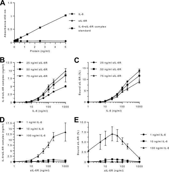
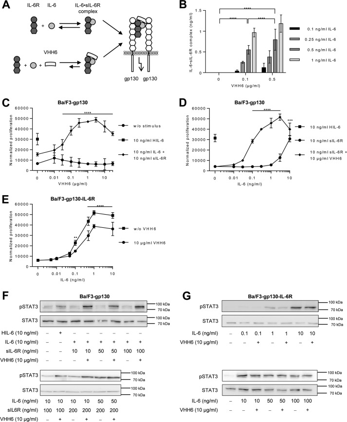
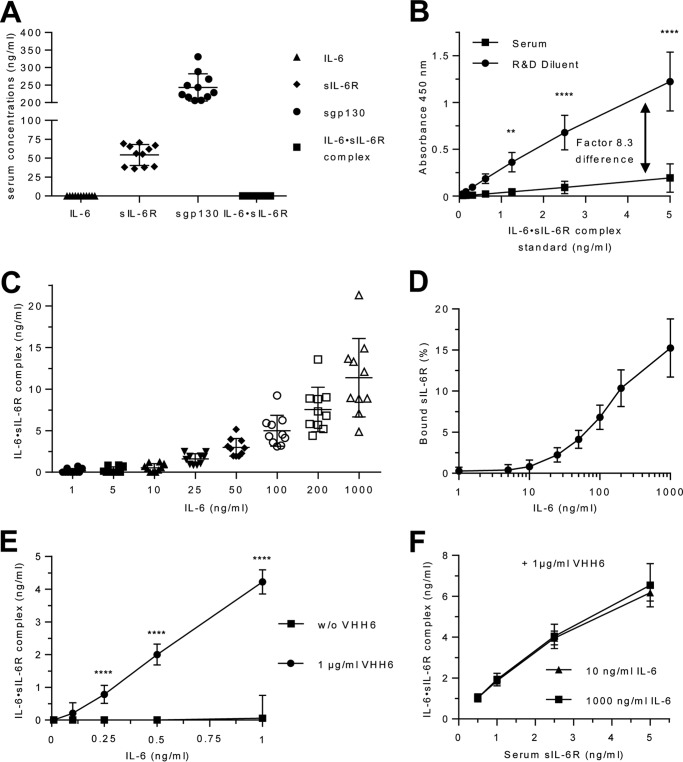
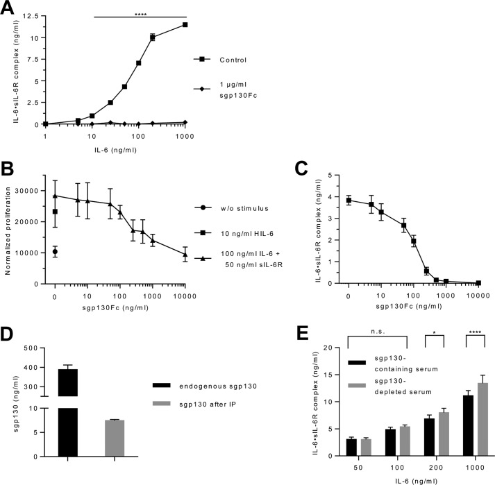
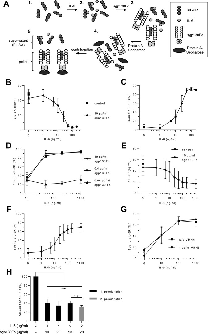
Interleukin (IL-)6 is the major pro-inflammatory cytokine within the IL-6 family. IL-6 signals via glycoprotein 130 (gp130) and the membrane-bound or soluble IL-6 receptor (IL-6R), referred to as classic or trans-signaling, respectively. Whereas inflammation triggers IL-6 expression, eventually rising to nanogram/ml serum levels, soluble IL-6R (sIL-6R) and soluble gp130 (sgp130) are constitutively present in the upper nanogram/ml range. Calculations based on intermolecular affinities have suggested that systemic IL-6 is immediately trapped in IL-6·sIL-6R and IL-6·sIL-6R·sgp130 complexes, indicating that sIL-6R and sgp130 constitute a buffer system that increases the serum half-life of IL-6 or restricts systemic IL-6 signaling. However, this scenario has not been experimentally validated. Here, we quantified IL-6·sIL-6R and IL-6·sIL-6R·sgp130 complexes over a wide concentration range. The amounts of IL-6 used in this study reflect concentrations found during active inflammatory events. Our results indicated that most IL-6 is free and not complexed with sIL-6R or sgp130, indicating that the level of endogenous sgp130 in the bloodstream is not sufficient to block IL-6 trans-signaling via sIL-6R. Importantly, addition of the single-domain antibody VHH6, which specifically stabilizes IL-6·sIL-6R complexes but did not bind to IL-6 or sIL-6R alone, drove free IL-6 into IL-6·sIL-6R complexes and boosted trans-signaling but not classic signaling, demonstrating that endogenous sIL-6R has at least the potential to form complexes with IL-6. Our findings indicate that even though high concentrations of sIL-6R and sgp130 are present in human serum, the relative ratio of free IL-6 to IL-6·sIL-6R allows for simultaneous classic and trans-signaling.

Keywords: Trans-signaling; classic signaling; cytokine; glycoprotein; interleukin 6 (IL-6); interleukin 6 receptor (IL6R); signal transduction.


Conflict of interest statement

G. H. W. is employed by CONARIS Research Institute AG (Kiel, Germany), which is commercially developing sgp130Fc proteins as therapeutics for inflammatory diseases

Figures

Figure 1.
Figure 1.
Quantification of IL-6·sIL-6R complexes. A, validation of the IL-6·sIL-6R ELISA was performed with recombinant IL-6 (0.078–5 ng/ml), sIL-6R (0.078–5 ng/ml), or the IL-6·sIL-6R complex standard (0.078–5 ng/ml). B, recombinant human IL-6 (1–1,000 ng/ml) and sIL-6R (25, 50, or 75 ng/ml) were mixed and IL-6·sIL-6R complexes were quantified by IL-6·sIL-6R ELISA. Combined data from three experiments are shown. Error bars represent the S.D. C, the percentages of sIL-6R present in IL-6·sIL-6R complexes were calculated from B. The calculation is based on the molecular masses of IL-6 (23.7 kDa) and sIL-6R (51.5 kDa), bound in the IL-6·sIL-6R complex (75.2 kDa, ratio = ∼1/3 IL-6 and 2/3 sIL-6R). For example, 5 ng/ml of IL-6/sIL-6R were detected, by combination of 100 ng/ml of IL-6 and 50 ng/ml of sIL-6R. Thus the complex consists of 3.42 ng/ml of sIL-6R and 1.58 ng/ml of IL-6. Consequently, 6.85% of the used sIL-6R and 1.57% of the used IL-6 were bound in the IL-6·sIL-6R complex. Error bars represent the S.D. D, recombinant human sIL-6R (1–1,000 ng/ml) and IL-6 (1, 10, or 100 ng/ml) were mixed and IL-6·sIL-6R complexes were quantified by IL-6·sIL-6R ELISA. Combined data were from three independent experiments. Error bars represent the S.D. E, the percentages of sIL-6R present in IL-6·sIL-6R complexes were calculated from D.
Figure 2.
Figure 2.
VHH6 promotes IL-6 trans-signaling and IL-6·sIL-6R complex formation. A, schematic illustration of VHH6 binding to IL-6·sIL-6R complexes. B, recombinant human IL-6 (0.1–1 ng/ml) and sIL-6R (50 ng/ml) plus increasing amounts of VHH6 (0.1 or 0.5 μg/ml) were mixed and the formed IL-6·sIL-6R complexes were quantified by IL-6·sIL-6R ELISA. Combined data were from three experiments. Error bars represent S.D. C, cellular proliferation of Ba/F3–gp130 cells. VHH6 (0.01–30 μg/ml) was titrated into a setup in which equal numbers of cells were cultured for 3 days without stimulus, with 10 ng/ml of Hyper-IL-6 (HIL-6; control representing 100% trans-signaling complex) or with a combination of 10 ng/ml of IL-6 and 10 ng/ml of sIL-6R. Proliferation was measured using the colorimetric CellTiter-Blue Cell Viability Assay. One representative experiment of three is shown. Error bars represent the S.D. D, cellular proliferation of Ba/F3-gp130 cells. IL-6 (0.01–10 ng/ml) was titrated into a setup in which equal numbers of cells were cultured for 3 days with sIL-6R (10 ng/ml) or with a combination of sIL-6R (10 ng/ml) and VHH6 (10 μg/ml) or HIL-6 alone (10 ng/ml). Proliferation was measured using the colorimetric CellTiter-Blue Cell Viability Assay. One representative experiment of three is shown. Error bars represent the S.D. E, cellular proliferation of Ba/F3-gp130-IL-6R cells. Equal numbers of cells were cultured for 3 days in the presence of HIL-6 (10 ng/ml) or VHH6 (10 μg/ml) and increasing concentrations of IL-6 (0.005–10 ng/ml). Proliferation was measured using the colorimetric CellTiter-Blue Cell Viability Assay. One representative experiment of three is shown. Error bars represent the S.D. F, analysis of STAT3 activation following trans-signaling. Ba/F3–gp130 cells were washed three times, starved, and stimulated with the indicated amounts of HIL-6, IL-6, sIL-6R, and VHH6 for 10 min. Cellular lysates were prepared, and equal amounts of total protein (50 μg/lane) were loaded on SDS gels, followed by immunoblotting using antibodies against phospho-STAT3 or STAT3 (loading control, same samples were used on separated gels). Western blotting data show one representative experiment of three. G, analysis of STAT3 activation following classic signaling. Ba/F3–gp130–IL-6R cells were washed three times, starved, and stimulated with the indicated amounts of IL-6 and VHH6 for 10 min. Cellular lysates were prepared, and equal amounts of total protein (50 μg/lane) were loaded on SDS gels, followed by immunoblotting using antibodies against phospho-STAT3 or STAT3 (loading control, same samples were used on separated gels). Western blotting data show one representative experiment of three.
Figure 3.
Figure 3.
Quantification of IL-6·sIL-6R complexes in human serum. A, serum level of IL-6, sIL-6R, sgp130, and IL-6·sIL-6R complexes from 10 human healthy volunteers were quantified by ELISA. Bars represent the mean (middle line) and the S.D. (upper and lower line). B, IL-6·IL-6R complex standard from R&D Systems was reconstituted either in the recommended diluent or in human serum. The calculation of the correction factor for IL-6·sIL-6R complex detection in serum (8.3 times) was performed with pooled data from three independent experiments. Error bars represent the S.D. C, recombinant human IL-6 (1–1,000 ng/ml) was titrated into serum samples from 10 human healthy volunteers, and the resulting IL-6·sIL-6R complexes were quantified by IL-6·sIL-6R complex ELISA. Bars represent the mean (middle line) and the S.D. (upper and lower line). D, the percentages of sIL-6R in IL-6·sIL-6R complexes were calculated from C. Error bars represent the S.D. E, recombinant human IL-6 (0.1–1 ng/ml) and VHH6 (0 or 1 μg/ml) were added to serum samples from 3 human healthy volunteers, and the resulting IL-6·sIL-6R complexes were quantified by IL-6·sIL-6R complex ELISA. Combined data were from three experiments. Error bars represent the S.D. F, recombinant human IL-6 (10 or 1,000 ng/ml), VHH6 (1 μg/ml), and endogenous sIL-6R (0.5, 1, 2.5, or 5 ng/ml) in the serum of three human healthy volunteers were mixed and IL-6·sIL-6R complex was quantified by IL-6·sIL-6R complex ELISA. Combined data from three experiments. Error bars represent the S.D.
Figure 4.
Figure 4.
Influence of sgp130Fc on the ELISA quantification of IL-6·sIL-6R complexes in human serum. A, recombinant human IL-6 (1–1,000 ng/ml), sIL-6R (50 ng/ml), and sgp130Fc (0 or 1 μg/ml) were mixed and the resulting IL-6·sIL-6R complexes were quantified by IL-6·sIL-6R complex ELISA. One representative experiment of three is shown. Error bars represent the S.D. B, cellular proliferation of Ba/F3-gp130 cells. Equal numbers of cells were cultured for 3 days in the presence of HIL-6 (10 ng/ml) or IL-6 (100 ng/ml) plus sIL-6R (50 ng/ml) and increasing concentrations of sgp130Fc (0.005–10 μg/ml). Proliferation was measured using the colorimetric CellTiter-Blue Cell Viability Assay. One representative experiment of three is shown. Error bars represent the S.D. C, recombinant human IL-6 (100 ng/ml), sIL-6R (50 ng/ml), and sgp130Fc (5–10,000 ng/ml) were mixed and the resulting IL-6·sIL-6R complexes were quantified by IL-6·sIL-6R complex ELISA. Combined data from three experiments. Error bars represent the S.D. D, endogenous sgp130 was removed from the serum of one healthy volunteer by immunoprecipitation with an anti-sgp130 antibody and Protein A-Sepharose. Endogenous sgp130 in the serum before and after precipitation was quantified by sgp130 ELISA. E, recombinant human IL-6 (50–1,000 ng/ml) was added to the endogenous sIL-6R in natural and sgp130-depleted serum of one human healthy volunteer and the resulting IL-6·sIL-6R complexes were quantified by IL-6·sIL-6R complex ELISA. Combined data were from three experiments. Error bars represent the S.D.
Figure 5.
Figure 5.
Quantification of IL-6·sIL-6R·sgp130Fc complexes. A, schematic illustration of the precipitation of IL-6·sIL-6R·sgp130Fc by Protein A-Sepharose beads and subsequent analysis of the remaining sIL-6R in the supernatant. B, recombinant human IL-6 (1–200 ng/ml), sIL-6R (50 ng/ml), and sgp130Fc (10 μg/ml) were mixed. The resulting IL-6·sIL-6R·sgp130Fc complexes were precipitated with Protein A-Sepharose and sIL-6R in the supernatant was quantified by ELISA. Combined data were from five experiments. Error bars represent the S.D. C, the percentages of sIL-6R in IL-6·sIL-6R·sgp130Fc complexes were calculated from B. Error bars represent the S.D. D, recombinant human IL-6 (10–1,000 ng/ml), sIL-6R (50 ng/ml), and sgp130Fc (0.04–10 μg/ml) were mixed. The resulting IL-6·sIL-6R·sgp130Fc complexes were precipitated with Protein A-Sepharose and sIL-6R was quantified in the supernatant by ELISA. Mean of combined data were from three experiments. Error bars represent the S.D. E, recombinant human IL-6 (1–1,000 ng/ml), endogenous (serum) sIL-6R, and sgp130Fc (10 μg/ml) were mixed. The resulting IL-6·sIL-6R·sgp130Fc complexes were precipitated with Protein A-Sepharose and sIL-6R was quantified in the supernatant by ELISA. Serum of 10 human healthy volunteers was used. Mean of combined data were from 10 measurements. Error bars represent the S.D. F, the percentages of endogenous (serum) sIL-6R in IL-6·sIL-6R·sgp130Fc complexes were calculated from E. Error bars represent the S.D. G, recombinant human IL-6 (1–1,000 ng/ml), endogenous (serum) sIL-6R, sgp130Fc (10 μg/ml), and VHH6 (0 or 1 μg/ml) were mixed. IL-6·sIL-6R·sgp130Fc complexes were precipitated with Protein A-Sepharose and sIL-6R was quantified in the supernatant by ELISA. Mean of combined data were from three experiments. Error bars represent the S.D. H, recombinant human IL-6 (1 or 2 μg/ml), endogenous (serum) sIL-6R and sgp130Fc (10 or 20 μg/ml) were mixed. The resulting IL-6·sIL-6R·sgp130Fc complexes were precipitated with Protein A-Sepharose and sIL-6R was quantified in the supernatant by ELISA. The precipitation and sIL-6R ELISA quantification was repeated after a second addition of IL-6 (2,000 ng/ml) and sgp130Fc (20 μg/ml). Mean of combined data were from three experiments. Error bars represent the S.D.

References

    1. Scheller J., Chalaris A., Schmidt-Arras D., and Rose-John S. (2011) The pro- and anti-inflammatory properties of the cytokine interleukin-6. Biochim. Biophys. Acta 1813, 878–888 10.1016/j.bbamcr.2011.01.034 - DOI - PubMed
    1. Garbers C., Hermanns H. M., Schaper F., Müller-Newen G., Grötzinger J., Rose-John S., and Scheller J. (2012) Plasticity and cross-talk of Interleukin 6-type cytokines. Cytokine Growth Factor Rev. 23, 85–97 10.1016/j.cytogfr.2012.04.001 - DOI - PubMed
    1. Jostock T., Mullberg J., Ozbek S., Atreya R., Blinn G., Voltz N., Fischer M., Neurath M. F., and Rose-John S. (2001) Soluble gp130 is the natural inhibitor of soluble interleukin-6 receptor transsignaling responses. Eur. J. Biochem. 268, 160–167 10.1046/j.1432-1327.2001.01867.x - DOI - PubMed
    1. Horiuchi S., Koyanagi Y., Zhou Y., Miyamoto H., Tanaka Y., Waki M., Matsumoto A., Yamamoto M., and Yamamoto N. (1994) Soluble interleukin-6 receptors released from T cell or granulocyte/macrophage cell lines and human peripheral blood mononuclear cells are generated through an alternative splicing mechanism. Eur. J. Immunol. 24, 1945–1948 10.1002/eji.1830240837 - DOI - PubMed
    1. Matthews V., Schuster B., Schütze S., Bussmeyer I., Ludwig A., Hundhausen C., Sadowski T., Saftig P., Hartmann D., Kallen K.-J., and Rose-John S. (2003) Cellular cholesterol depletion triggers shedding of the human interleukin-6 receptor by ADAM10 and ADAM17 (TACE). J. Biol. Chem. 278, 38829–38839 10.1074/jbc.M210584200 - DOI - PubMed

Publication types

MeSH terms