. 2018 Apr 1;9(4):428.
doi: 10.1038/s41419-018-0435-y.

PFKFB3 blockade inhibits hepatocellular carcinoma growth by impairing DNA repair through AKT

Affiliations

PFKFB3 blockade inhibits hepatocellular carcinoma growth by impairing DNA repair through AKT

Wen-Kai Shi et al. Cell Death Dis. .

Abstract

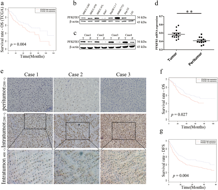
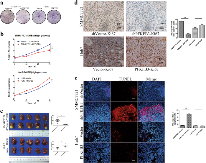
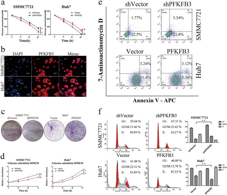
Overexpression of 6-phosphofructo-2-kinase/fructose-2,6-biphosphatase 3 (PFKFB3), a key molecule of glucose metabolism in cytoplasm, has been found in various tumors. Emerging evidence has suggested that PFKFB3 is also located in the nucleus and apparent in regulatory functions other than glycolysis. In this study, we found that PFKFB3 expression is associated with hepatocellular carcinoma (HCC) growth and located mainly in the nucleus of tumor cells. PFKFB3 overexpression was associated with large tumor size (p = 0.04) and poor survival of patients with HCC (p = 0.027). Knockdown of PFKFB3 inhibited HCC growth, not only by reducing glucose consumption but also by damaging the DNA repair function, leading to G2/M phase arrest and apoptosis. In animal studies, overexpression of PFKFB3 is associated with increased tumor growth. Mechanistically, PFKFB3 silencing decreased AKT phosphorylation and reduced the expression of ERCC1, which is an important DNA repair protein. Moreover, PFK15, a selective PFKFB3 inhibitor, significantly inhibited tumor growth in a xenograft model of human HCC. PFKFB3 is a potential novel target in the treatment of HCC.


Conflict of interest statement

The authors declare that they have no conflict of interest.

Figures

Fig. 1
Fig. 1. PFKFB3 overexpressed in liver cancer cells and correlated with poor prognosis in HCC patients.
a Kaplan–Meier analysis for overall survival (OS) of patients based on the data of TCGA. The OS of the PFKFB3 high expression group was worse than that of the PFKFB3 low expression group (p = 0.004). b Western blot analysis of PFKFB3 protein expression in liver cancer cell lines and normal hepatocyte cell line (L02). The PFKFB3 expressed higher in liver cancer cell lines. c Western blot analysis of PFKFB3 protein expression in paired human hepatocellular carcinoma (HCC) and peritumor tissue (T = Tumor tissue, P = Peritumor tissue). The PFKFB3 protein expressed higher in tumor tissues. d qRT-PCR expression analysis of PFKFB3 mRNA in paired human HCC and peritumor tissue (n = 12). The PFKFB3 mRNA expressed higher in tumor tissues. e Immunohistochemistry (IHC) staining of paired human HCC and peritumor tissue. PFKFB3 protein expressed higher in tumor tissues and located mainly in nucleus in human liver cancer tissue. f Kaplan–Meier analysis for OS based on the data of our center. The OS (p = 0.027) of the high expression group were worse than that of the PFKFB3 low expression group. g Kaplan–Meier analysis for disease-free survival (DFS) based on the data of our center. The DFS (p = 0.004) of the high expression group were worse than that of the PFKFB3 low expression group. **p < 0.01
Fig. 2
Fig. 2. PFKFB3 expression in HCC cells promoted tumor growth in vitro and in vivo.
a Clone formation of SMMC7721-shPFKFB3 cells and Huh7-PFKFB3 cells compared with their vector control. PFKFB3 promoted the cells clone formation in vitro. b CCK8 assay for cell proliferation of SMMC7721-shPFKFB3 cells and Huh7-PFKFB3 cells compared with their vector control. PFKFB3 promoted the liver cancer cells proliferation in vitro. c Comparison of tumor sizes for the SMMC7721-shPFKFB3 cells group and SMMC7721-shVector cells group (45.3 ± 14.9 mm3 vs. 201.9 ± 88.6 mm3; p = 0.02), and comparison of tumor sizes for the Huh7-PFKFB3 cells group and Huh7-Vector cells group (825.6 ± 217.9 mm3 vs. 467.8 ± 221.9 mm3; p = 0.033). d Representative immunohistochemistry of liver cancer for the expression of Ki67 from Balb/c nu/nu mice orthotopically implanted with SMMC7721 or Huh7 cells. Ki67 expressed higher in PFKFB3 high expression tumor. (Magnification ×200). e Representative TUNEL fluorescence of liver cancer from Balb/c nu/nu mice orthotopically implanted with SMMC7721 or Huh7 cells. The apoptosis rate of cells was higher in PFKFB3 low expression tumor. (Magnification ×200). *p < 0.05, **p < 0.01
Fig. 3
Fig. 3. PFKFB3 inhibition led to G2/M phase arrest and apoptosis of HCC cells.
a Glucose consumption of different PFKFB3 expressions of SMMC7721 and Huh7 cells. High expression cells consumoted more glucose. b Immunofluorescence stain of PFKFB3 in SMMC7721 and Huh7 cells. PFKFB3 located both in nucleus and cytoplasm. c Clone formation of SMMC7721-shPFKFB3 cells and Huh7-PFKFB3 cells compared with their vector control in glucose substitute medium. PFKFB3 promoted the cells clone formation eliminating the effect of glucose in vitro. d CCK8 assay for cell proliferation of SMMC7721-shPFKFB3 cells and Huh7-PFKFB3 cells compared with their vector control in glucose substitute medium. PFKFB3 promoted the liver cancer cells proliferation eliminating the effect of glucose in vitro. e Flow cell apoptosis detection for cell apoptosis rates of SMMC7721-shPFKFB3 cells and Huh7-PFKFB3 cells compared with their vector control in glucose substitute medium. PFKFB3 decreased the apoptosis rate of liver cancer cells eliminating the effect of glucose in vitro. f Flow cytometry cycle detection of the cell cycle ratio of SMMC7721-shPFKFB3 cells and Huh7-PFKFB3 cells compared with their vector control in glucose substitute medium. PFKFB3 knockdown increased the proportion of G2/M phase. **p < 0.01
Fig. 4
Fig. 4. PFKFB3 knockdown inhibited DNA repair via the AKT/ERCC1 pathway.
a Comet assay for DNA damage of SMMC7721-shPFKFB3 and SMMC7721-shVector. In SMMC7721-shPFKFB3 cells, DNA damage presented more compared with SMMC7721-shVector cells. b The ATM/Chk1/cdc25C signaling pathway-associated proteins were detected by Western blot in SMMC7721-shPFKFB3 and Huh7-PFKFB3 compared with their vector control. c Immunoprecipitation assay was conducted for SMMC7721 cells, and IgG was used as a control. AKT was immunoprecipitated by anti-PFKFB3 antibody. d The PFKFB3, AKT, pAKT, and ERCC1 were detected by Western blot in SMMC7721-shPFKFB3 and Huh7-PFKFB3 compared with their vector control. e Inhibited AKT phosphorylated by MK-2206 in Huh7-PFKFB3 cells. PFKFB3 expression was not increased but ERCC1 expression was decreased in Huh7-PFKFB3 cells, and the ATM/Chk1/cdc25C signaling pathway had no significant change. **p < 0.01
Fig. 5
Fig. 5. PFKFB3 inhibitor PFK15 inhibits HCC growth in vivo and in vitro.
a CCK8 assay for cell proliferation of SMMC7721 cells and Huh7 cells treated with PFK15 in a dose and time series. PFK15 inhibited proliferation of SMMC7721 and Huh7 in a dose-dependent and time-dependent manner. b Flow cell apoptosis detection of the cell apoptosis rate of SMMC7721 cells and Huh7 cells treated with PFK15 in a dose and time series. PFK15 increased the apoptosis rate of SMMC7721 and Huh7 in a dose-dependent and time-dependent manner. c Flow cytometry cycle detection of the cell cycle ratio of SMMC7721 cells and Huh7 cells treated with PFK15 in a dose and time series. PFK15 induced G2/M arrest of SMMC7721 and Huh7 in a dose-dependent and time-dependent manner. d Comet assay for DNA damage of SMMC7721 cells and Huh7 cells treated with PFK15 (24 h). PFK15 led to DNA damage in vitro. e Comparison of tumor sizes of the SMMC7721-control group and SMMC7721-PFK15 group (1062.2 ± 578.2 mm3 vs. 215.9 ± 104.8 mm3; p = 0.03), and comparison of tumor sizes of the Huh7-control group and Huh7-PFK15 group (1628.4 ± 495.5 mm3 vs. 475.4 ± 222.9 mm3; p = 0.004). PFK15 treatment delayed tumor growth in both SMMC7721 and Huh7 tumor models. f Representative immunohistochemistry of liver cancer showing the expressions of Ki67 and ERCC1 from Balb/c nu/nu mice orthotopically implanted with SMMC7721 or Huh7 cells. PFK15 treatment decreased the expression of ERCC1 and Ki67.(Magnification ×200). g Representative TUNEL fluorescence of liver cancer from Balb/c nu/nu mice orthotopically implanted with SMMC7721 or Huh7 cells. PFK15 treatment increased the apoptosis rate of tumor.(Magnification ×200). **p < 0.01

Similar articles

Cited by

References

    1. Llovet JM, et al. Hepatocellular carcinoma. Nat. Rev. Dis. Prim. 2016;2:16018. doi: 10.1038/nrdp.2016.18. - DOI - PubMed
    1. Llovet JM, et al. Arterial embolisation or chemoembolisation versus symptomatic treatment in patients with unresectable hepatocellular carcinoma: a randomised controlled trial. Lancet. 2002;359:1734–1739. doi: 10.1016/S0140-6736(02)08649-X. - DOI - PubMed
    1. Llovet JM, et al. Sorafenib in advanced hepatocellular carcinoma. N. Engl. J. Med. 2008;359:378–390. doi: 10.1056/NEJMoa0708857. - DOI - PubMed
    1. Hanahan D, Weinberg RA. Hallmarks of cancer: the next generation. Cell. 2011;144:646–674. doi: 10.1016/j.cell.2011.02.013. - DOI - PubMed
    1. Warburg O. On the origin of cancer cells. Science. 1956;123:309–314. doi: 10.1126/science.123.3191.309. - DOI - PubMed

Publication types

MeSH terms