

Modeling Environmentally-Induced Motor Neuron Degeneration in Zebrafish
- PMID: 29559645
- PMCID: PMC5861069
- DOI: 10.1038/s41598-018-23018-w
Modeling Environmentally-Induced Motor Neuron Degeneration in Zebrafish
Abstract
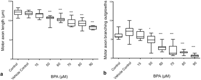
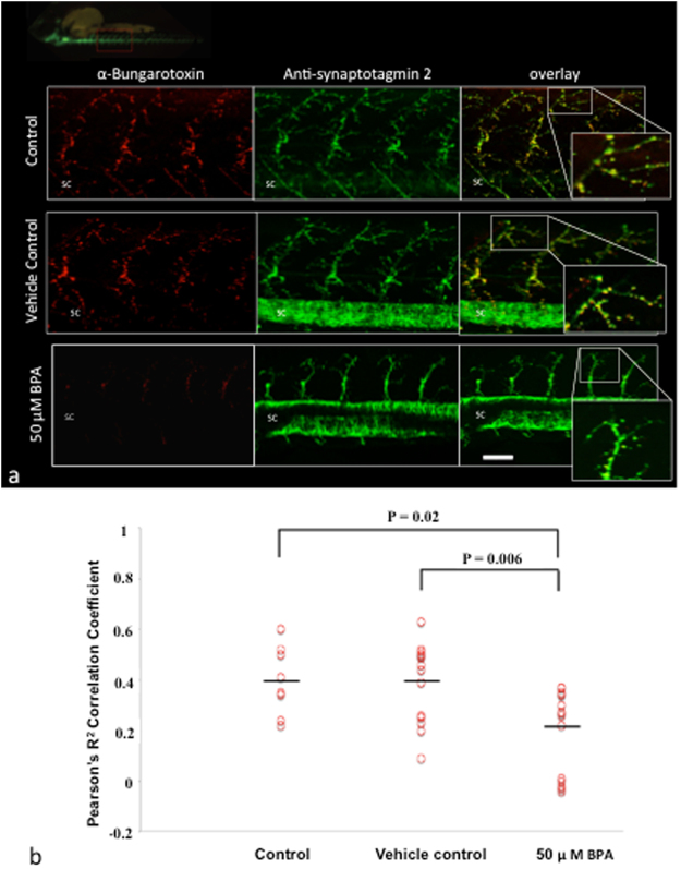
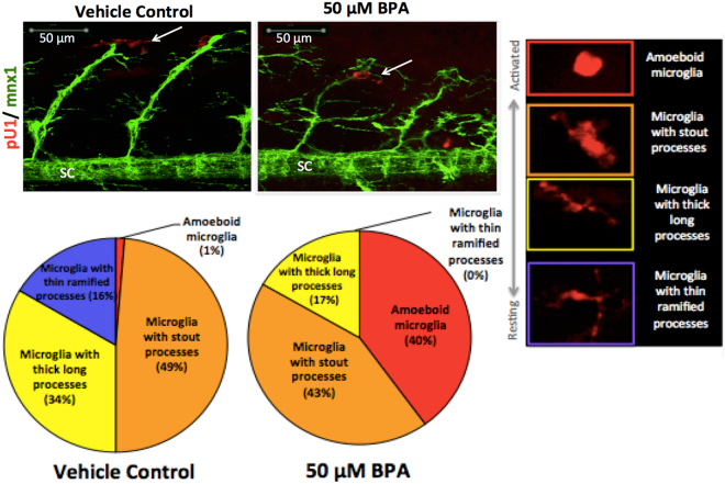
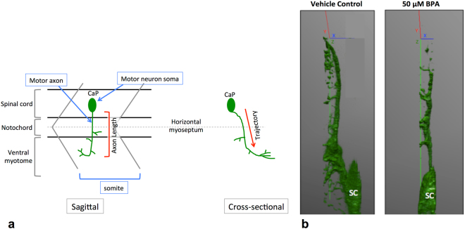
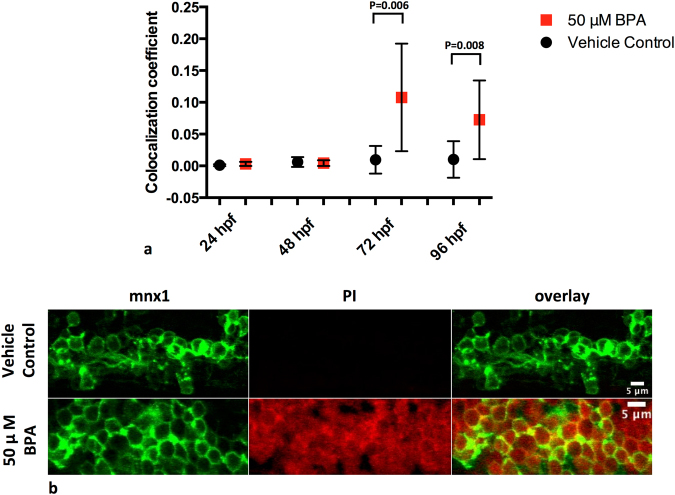
Zebrafish have been used to investigate motor neuron degeneration, including as a model system to examine the pathogenesis of amyotrophic lateral sclerosis (ALS). The use of zebrafish for this purpose has some advantages over other in vivo model systems. In the current paper, we show that bisphenol A (BPA) exposure in zebrafish embryos results in motor neuron degeneration with affected motor function, reduced motor axon length and branching, reduced neuromuscular junction integrity, motor neuron cell death and the presence of activated microglia. In zebrafish, motor axon length is the conventional method for estimating motor neuron degeneration, yet this measurement has not been confirmed as a valid surrogate marker. We also show that reduced motor axon length as measured from the sagittal plane is correlated with increased motor neuron cell death. Our preliminary timeline studies suggest that axonopathy precedes motor cell death. This outcome may have implications for early phase treatments of motor neuron degeneration.
Conflict of interest statement
CAS is a founding member of Neurodyn Inc.
Figures
Similar articles
-
Altered terminal Schwann cell morphology precedes denervation in SOD1 mice.Exp Neurol. 2016 Jan;275 Pt 1(0 1):172-81. doi: 10.1016/j.expneurol.2015.09.014. Epub 2015 Sep 26. Exp Neurol. 2016. PMID: 26416261 Free PMC article.
-
Conditional Overexpression of rtn4al in Muscle of Adult Zebrafish Displays Defects Similar to Human Amyotrophic Lateral Sclerosis.Mar Biotechnol (NY). 2019 Feb;21(1):52-64. doi: 10.1007/s10126-018-9857-x. Epub 2018 Nov 15. Mar Biotechnol (NY). 2019. PMID: 30443836
-
Nucleo-cytoplasmic transport of TDP-43 studied in real time: impaired microglia function leads to axonal spreading of TDP-43 in degenerating motor neurons.Acta Neuropathol. 2018 Sep;136(3):445-459. doi: 10.1007/s00401-018-1875-2. Epub 2018 Jun 25. Acta Neuropathol. 2018. PMID: 29943193 Free PMC article.
-
Axonal degeneration in motor neuron disease.Neurodegener Dis. 2007;4(6):431-42. doi: 10.1159/000107704. Epub 2007 Oct 9. Neurodegener Dis. 2007. PMID: 17934327 Review.
-
Neuromuscular junction destruction during amyotrophic lateral sclerosis: insights from transgenic models.Curr Opin Pharmacol. 2009 Jun;9(3):341-6. doi: 10.1016/j.coph.2009.03.007. Epub 2009 Apr 20. Curr Opin Pharmacol. 2009. PMID: 19386549 Review.
Cited by
-
Omics Approach to Axonal Dysfunction of Motor Neurons in Amyotrophic Lateral Sclerosis (ALS).Front Neurosci. 2020 Mar 25;14:194. doi: 10.3389/fnins.2020.00194. eCollection 2020. Front Neurosci. 2020. PMID: 32269505 Free PMC article. Review.
-
Does Bisphenol A Confer Risk of Neurodevelopmental Disorders? What We Have Learned from Developmental Neurotoxicity Studies in Animal Models.Int J Mol Sci. 2022 Mar 7;23(5):2894. doi: 10.3390/ijms23052894. Int J Mol Sci. 2022. PMID: 35270035 Free PMC article. Review.
-
Neuronal branching is increasingly asymmetric near synapses, potentially enabling plasticity while minimizing energy dissipation and conduction time.J R Soc Interface. 2023 Sep;20(206):20230265. doi: 10.1098/rsif.2023.0265. Epub 2023 Sep 6. J R Soc Interface. 2023. PMID: 37669695 Free PMC article.
-
Zebrafish as a model organism for neurodegenerative disease.Front Mol Neurosci. 2022 Oct 13;15:940484. doi: 10.3389/fnmol.2022.940484. eCollection 2022. Front Mol Neurosci. 2022. PMID: 36311026 Free PMC article. Review.
-
Mitochondrial Dyshomeostasis as an Early Hallmark and a Therapeutic Target in Amyotrophic Lateral Sclerosis.Int J Mol Sci. 2023 Nov 27;24(23):16833. doi: 10.3390/ijms242316833. Int J Mol Sci. 2023. PMID: 38069154 Free PMC article. Review.
References
MeSH terms
Substances
LinkOut - more resources
Full Text Sources
Other Literature Sources
Molecular Biology Databases
Miscellaneous
