Review
. 2018 Mar 21;14(4):214-228.
doi: 10.1038/nrrheum.2018.31.

Type I interferon in rheumatic diseases

Affiliations
Review

Type I interferon in rheumatic diseases

Theresa L Wampler Muskardin et al. Nat Rev Rheumatol. .

Abstract

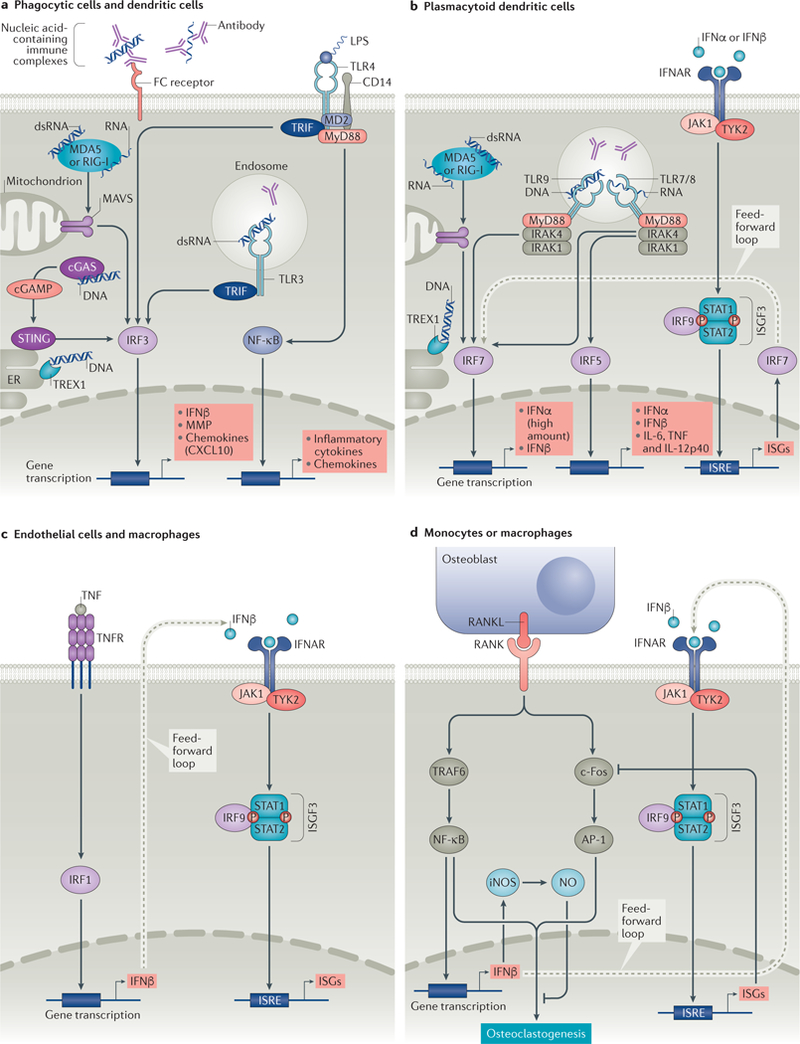
The type I interferon pathway has been implicated in the pathogenesis of a number of rheumatic diseases, including systemic lupus erythematosus, Sjögren syndrome, myositis, systemic sclerosis, and rheumatoid arthritis. In normal immune responses, type I interferons have a critical role in the defence against viruses, yet in many rheumatic diseases, large subgroups of patients demonstrate persistent activation of the type I interferon pathway. Genetic variations in type I interferon-related genes are risk factors for some rheumatic diseases, and can explain some of the heterogeneity in type I interferon responses seen between patients within a given disease. Inappropriate activation of the immune response via Toll-like receptors and other nucleic acid sensors also contributes to the dysregulation of the type I interferon pathway in a number of rheumatic diseases. Theoretically, differences in type I interferon activity between patients might predict response to immune-based therapies, as has been demonstrated for rheumatoid arthritis. A number of type I interferon and type I interferon pathway blocking therapies are currently in clinical trials, the results of which are promising thus far. This Review provides an overview of the many ways in which the type I interferon system affects rheumatic diseases.


Figures

Fig. 1 |
Fig. 1 |. Major pathways of induction of type I interferon production in different cell lineages.
a | In phagocytes and dendritic cells, stimulation of surface Toll-likereceptor 4 (TLR4) by lipopolysaccharide (LPS) or endosomal TLR3 by double-stranded RNA (dsRNA) results in activation of interferon regulatory factor 3 (IRF3) via a TIR domain-containing adaptor molecule 1 (TICAM-1, also known as TRIF)-dependent pathway, and nuclear factor-κB (NF-κB) via myeloid differentiation primary response protein (MyD88). Activation of cytosolic nucleic acid sensors (melanoma differentiation-associated protein 5 (MDA5) or retinoic acid inducible gene 1(RIG-I) by RNA, or stimulator of interferon genes protein (STING) by DNA (via cyclic GMP-AMP synthase (cGAS)) also prompt activation of IRF3. IRF3 translocates to the nucleus and induces transcription of IFNβ. b | In plasmacytoid dendritic cells (pDCs), activation of endosomal TLR7 or TLR8 by RNA results in activation of IRF7 and/or IRF5. Activation of endosomal TLR9 by DNA or of cytosolic sensors MDA5 or RIG-I by RNA results in activation of IRF7. IRF7 translocates to the nucleus, where it induces transcription of type I interferons. Translocation of IRF5 to the nucleus culminates in transcription of type I interferons and pro-inflammatory cytokines. In pDCs, binding of type I interferon to the type I interferon receptor (IFNAR) results in activation of the canonical Janus kinase (JAK)–signal transducer and activator of transcription (STAT) pathway that results in transcription of type I interferon stimulated genes (ISGs). ISGs include IRF7, which provides a feed-forward mechanism for production of more type I interferon. c | In macrophages and endothelial cells, TNF induces IFNβ via IRF1 and can induce an IFNβ autocrine loop that acts in synergy with canonical TNF signals to induce sustained expression of inflammatory genes and delayed expression of STAT1-dependent ISGs that prime cells for enhanced responses to subsequent challenge. d | Receptor activator of nuclear factor-κB (RANK)–RANK ligand (RANKL) interaction activates TNF receptor-associated factor 6 (TRAF6) and c-Fos pathways. TRAF6 activation results in induction of NFκB. c-Fos, together with activator protein 1 (AP-1) leads to a cascade that promotes osteoclastogenesis. NF-κB and c-Fos stimulate production of IFNβ. IFNβ promotes transcription of genes that inhibit c-Fos activity and results in the induction of nitric oxide (NO), which inhibits osteoclastogenesis. cGAMP, cyclic GMP-AMP; CXCL10, CXC-chemokine 10; ER, endoplasmic reticulum; iNOS, inducible nitric oxide synthase; IRAK, interleukin-1 receptor-associated kinase; ISGF3, interferon-stimulated gene factor 3; ISRE, interferon-sensitive response element; MAVS, mitochondrial antiviral-signalling protein; MD2, myeloid differentiation 2; MMP, matrix metalloproteinase; TNFR, TNF receptor; TYK2, tyrosine kinase 2; TREX1, three-prime repair exonuclease 1.

Similar articles

Cited by

References

    1. Rice G et al. Clinical and molecular phenotype of Aicardi-Goutieres syndrome. Am. J. Hum. Genet 81, 713–725 (2007). - PMC - PubMed
    1. Baechler EC et al. Interferon-inducible gene expression signature in peripheral blood cells of patients with severe lupus. Proc. Natl Acad. Sci. USA 100, 2610–2615 (2003). - PMC - PubMed
    1. Bennett L et al. Interferon and granulopoiesis signatures in systemic lupus erythematosus blood. J. Exp. Med 197, 711–723 (2003). - PMC - PubMed
    1. Crow MK & Wohlgemuth J Microarray analysis of gene expression in lupus. Arthritis Res. Ther 5, 279–287 (2003). - PMC - PubMed
    1. Kariuki SN et al. Genetic analysis of the pathogenic molecular sub-phenotype interferon-α identifies multiple novel loci involved in systemic lupus erythematosus. Genes Immun 16, 15–23 (2015). - PMC - PubMed

Publication types

MeSH terms