. 2018 Jun;37(25):3456-3470.
doi: 10.1038/s41388-018-0221-4. Epub 2018 Mar 21.

Transmembrane TNF-alpha promotes chemoresistance in breast cancer cells

Affiliations

Transmembrane TNF-alpha promotes chemoresistance in breast cancer cells

Zunyue Zhang et al. Oncogene. 2018 Jun.

Abstract

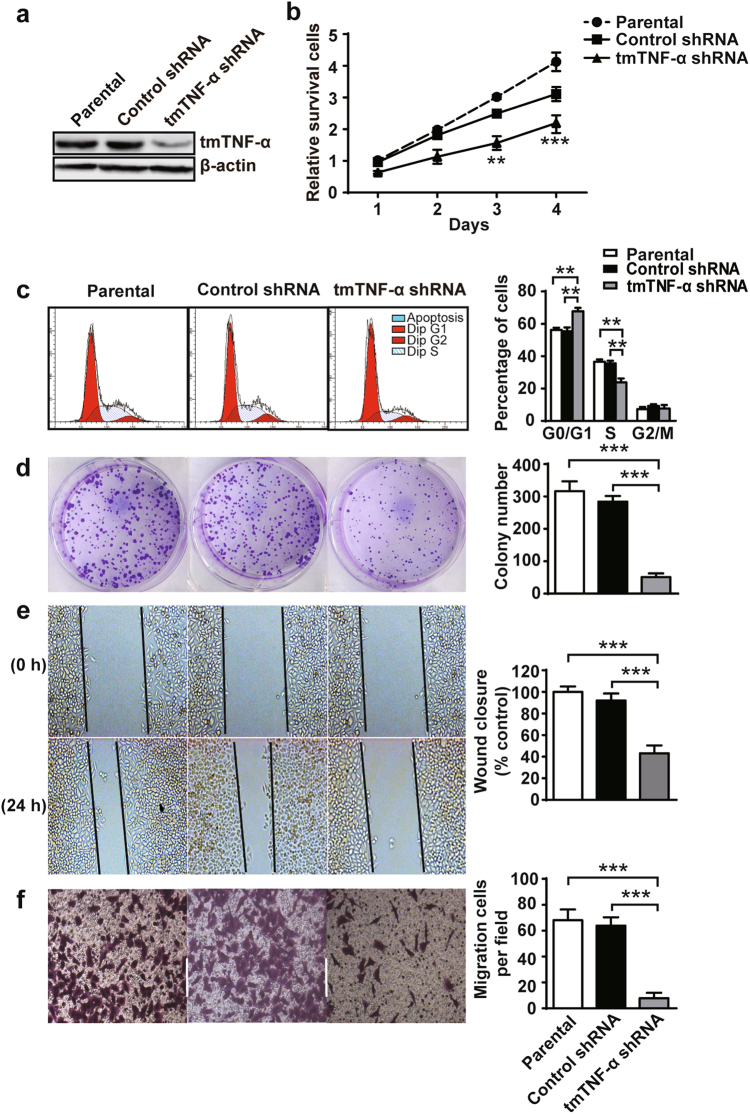
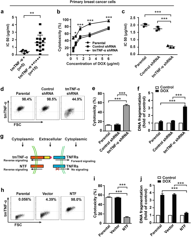
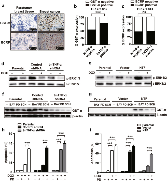
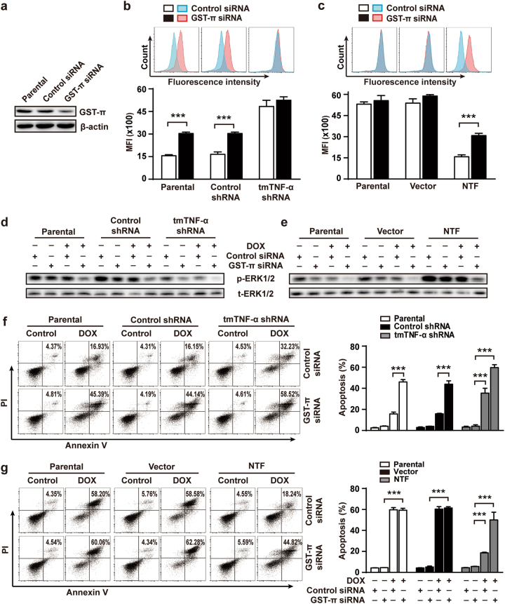
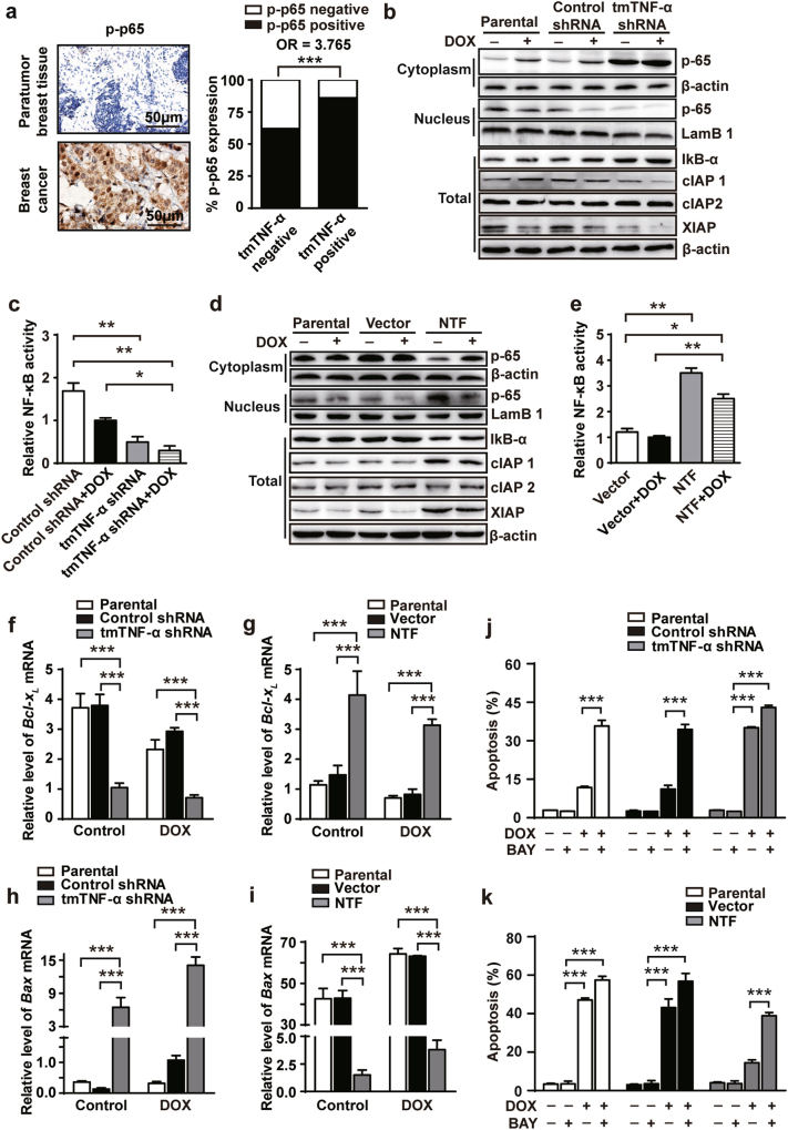
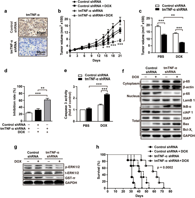
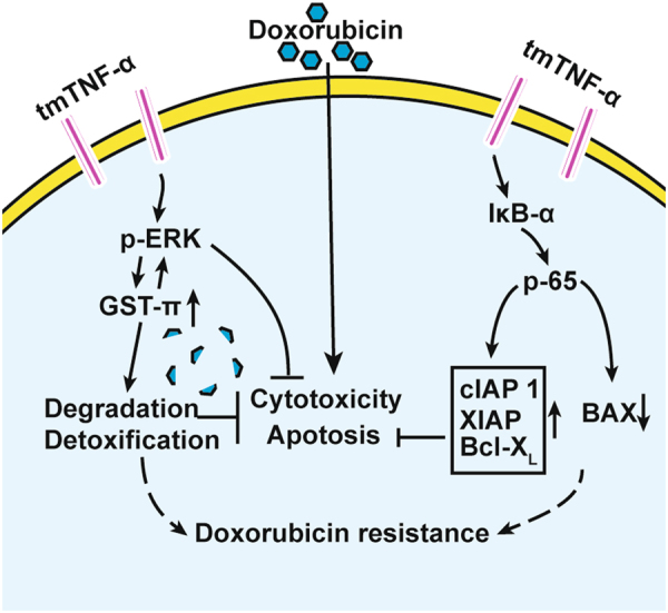
Chemoresistance remains a major obstacle to successful treatment of breast cancer. Although soluble tumor necrosis factor-α (sTNF-α) has been implicated in mediating drug-resistance in human cancers, whether transmembrane tumor necrosis factor-α (tmTNF-α) plays a role in chemoresistance remains unclear. Here we found that over 50% of studied patients expressed tmTNF-α at high levels in breast cancer tissues and tmTNF-α expression positively correlated with resistance to anthracycline chemotherapy. Alteration of tmTNF-α expression changed the sensitivity of primary human breast cancer cells and breast cancer cell lines to doxorubicin (DOX). Overexpression of N-terminal fragment (NTF) of tmTNF-α, a mutant form with intact intracellular domain of tmTNF-α to transmit reverse signals, induced DOX-resistance. Mechanistically, the tmTNF-α/NTF-ERK-GST-π axis and tmTNF-α/NTF-NF-κB-mediated anti-apoptotic functions were required for tmTNF-α-induced DOX-resistance. In a xenograft mouse model, the combination of tmTNF-α suppression with chemotherapy significantly enhanced the efficacy of DOX. Our data indicate that tmTNF-α mediates DOX-resistance through reverse signaling and targeting tmTNF-α may be beneficial for the treatment of DOX-resistant breast cancer.


Conflict of interest statement

The authors declare that they have no conflict of interest.

Figures

Fig. 1
Fig. 1
tmTNF-α promotes proliferation, clonogenicity, migration and invasion of MDA-MB-231. MDA-MB-231 breast cancer cells were stably transfected with control or tmTNF-α shRNA. a tmTNF-α expression determined by Western blot in parental or shRNA-transfected MDA-MB-231 cells. b Cell proliferation was detected after incubation for indicated time points by a CCK8 assay. c Quantification of DNA content by flow cytometry for analysis of cell cycle following propodium iodide staining. d Clonogenicity of parental or shRNA-transfected MDA-MB-231 cells was detected by colony formation assay after a 24-h culture. e Cell migration was evaluated using a wound-healing assay after 0 or 24-h culture. f Number of invasive cells cross through matrigel and membrane porous over 24 h. cf Representative images on the left, and the quantitative data on the right are presented as mean ± SEM of three independent experiments. **p < 0.01, ***p < 0.001
Fig. 2
Fig. 2
tmTNF-α is required for DOX-resistance of breast cancer. a Primary human breast cancer cells were isolated from patients with low (n = 5) or high (n = 15) expression of tmTNF-α and incubated with DOX for 72 h. IC50 values of DOX-mediated cytotoxicity was determined by ATP-TCA. b, c Primary breast cancer cells from three patients were transfected with control or tmTNF-α shRNA for 48 h. DOX was added in indicated concentrations and incubated for 72 h. b Dose dependent DOX-induced cytotoxicity determined by ATP-TCA. c IC50 values of DOX to primary cancer cells or those transfected with shRNA. d FACS analysis of tmTNF-α expression in parental or shRNA stably transfected MDA-MB-231 cells. e, f Cytotoxicity and DNA fragmentation of parental or shRNA-transfected MDA-MB-231 cells treated with 3 μM DOX for 24 h detected by MTT assay and ELISA, respectively. g Scheme of NTF-tmTNF-α-mediated reverse signaling without delivering forward signaling. h FACS analysis of NTF expression in parental, or empty vector or NTF-transfected MCF-7 cells. i and j Cytotoxicity and DNA fragmentation of parental or NTF-transfected MCF-7 cells treated with 3 μM DOX for 24 h. Data are represented as mean ± SEM of three independent experiments. *p < 0.05, **p < 0.01, ***p < 0.001
Fig. 3
Fig. 3
ERK-GST-π is involved in tmTNF-α-mediated DOX-resistance. a Representative IHC images of GST-π and BCRP expression in paratumor and breast cancer tumor sections. b, c Histogram showing co-expression rate of GST-π or BCRP with tmTNF-α in tumor tissues of 105 patients. d, e Immunoblotting analysis of phosphorylation levels of ERK1/2 in parental or shRNA-transfected MDA-MB-231 cells (d) and in parental or NTF-transfected MCF-7 cells (e) after treatment with or without 3 μM DOX for 24 h. f, g Immunoblotting analysis of GST-π expression in parental or shRNAs-transfected MDA-MB-231 cells and in parental or NTF-transfected MCF-7 cells treated with 10 μM BAY117082 (BAY), 10 μM PD98059 (PD) or 1 μM SCH772984 (SCH) for 24 h. h, i A 24-h DOX (3 μM)-induced apoptosis in parental or shRNA-transfected MDA-MB-231 cells and in parental or NTF-transfected MCF-7 cells pretreated with 10 μM PD98059 for 30 min using Annexin V/PI staining. Data are represented as mean ± SEM of three independent experiments. ***p < 0.001
Fig. 4
Fig. 4
Knockdown of GST-π expression blocked tmTNF-α-mediated DOX-resistance. Parental or shRNAs-transfected MDA-MB-231 cells and parental or NTF-transfected MCF-7 cells were transfected with 50 μM control or GST-π siRNA for 48 h and then treated with DOX (3 μM) for another 24 h. a Immunoblotting analysis of GST-π expression in MDA-MB-231 cells. b, c DOX fluorescence intensity was determined by FACS. d, e Immunoblotting analysis of phosphorylation levels of ERK1/2. f, g The apoptosis was determined by PI/Annexin V staining. Representative cytogram on the right and quantitative data on the left. Data are represented as mean ± SEM of three independent experiments. ***p < 0.001
Fig. 5
Fig. 5
NF-κB is required for tmTNF-α-mediated DOX-resistance. a Representative IHC images of phosphorylation of NF-κB p65 in paratumor and breast cancer tumors, and histogram showing co-expression rate of phosphorylated NF-κB p65 and tmTNF-α in tumors of 105 patients. (b-i) Parental or shRNA-transfected MDA-MB-231 cells and parental or NTF-transfected MCF-7 cells were treated with or without 3 μM DOX for 24 h. (b and d) Representative immunoblotting of levels of IκB-α, cIAP1, cIAP2, and XIAP in total protein and translocation of NF-κB p65 from cytoplasmic fraction to nuclear fraction. LamB1 or β-actin served as a nuclear protein or a cytoplasmic/total protein loading control. c, e NF-κB activity was detected by ELISA. fi Real-time PCR analysis of Bcl-XL (f and g) and BAX (h and i). j, k A 24 h-DOX (3 μM)-induced apoptosis was determined in parental or shRNA-transfected MDA-MB-231 cells (j) and in parental or NTF-transfected MCF-7 cells (k) pretreated with 10 μM BAY117082 (BAY) for 30 min using Annexin V/PI staining. Data are represented as mean ± SEM of three independent experiments. *p < 0.05, **p < 0.01, ***p < 0.001
Fig. 6
Fig. 6
Knockdown of tmTNF-α expression enhances the therapeutic efficacy of DOX in vivo. 1 × 106 MDA-MB-231 cells transfected with control or tmTNF-α shRNA were injected into mammary fat pad of nude mice. When tumors reached 100 mm3, the mice were treated intraperitoneally with DOX (4 mg/kg) or PBS, once a week for three weeks (n = 6 each group). a Representative images of tmTNF-α expression in tumor tissue section detected by IHC. b Growth curves of tumors after tumor cell inoculation. c Tumor sizes at the end point (day 21) of experiments. d Inhibition rate of tumor growth. e Caspase 3 activity in tumor tissues at day 21. f Immunoblotting of levels of IκB-а, cIAP1, cIAP2, XIAP, BAX, and Bcl-XL in total protein and translocation of NF-κB p65 from cytoplasmic fraction to nuclear fraction of tumor tissues. g Immunoblotting of levels of phosphorylation of ERK and GST-π in tumor tissues. h Kaplan–Meier survival curves of mice after tumor cell inoculation (n = 6). Data are represented as mean ± SEM. *p < 0.05, **p < 0.01, ***p < 0.001
Fig. 7
Fig. 7
tmTNF-α induces DOX-resistance of breast cancer cells via its reverse signaling. tmTNF-α can be overexpressed in breast cancer cells and tends to induce chemoresistance. tmTNF-α constitutively activates both ERK and NF-κB pathways through reverse signaling. tmTNF-α promotes GST-π expression via ERK pathway, leading to degradation and detoxification of DOX in tumor cells, and tmTNF-α-induced GST-π in turn further activates ERK pathway, forming a positive feedback to promote DOX-resistance. tmTNF-α can also upregulate antiapoptotic molecules via NF-κB pathway and downregulate proapoptotic molecule, resulting in survival and anti-apoptosis of tumor cells. Targeting tmTNF-α may sensitize tumor cells to chemotherapy

Similar articles

Cited by

References

    1. Arcamone F, Cassinelli G, Fantini G, Grein A, Orezzi P, Pol C, et al. Adriamycin, 14-hydroxydaunomycin, a new antitumor antibiotic from S. peucetius var. caesius. Reprinted from Biotechnology and Bioengineering, Vol. XI, Issue 6, Pages 1101–1110 (1969) Biotechnol Bioeng. 2000;67:704–13. doi: 10.1002/(SICI)1097-0290(20000320)67:6<704::AID-BIT8>3.0.CO;2-L. - DOI - PubMed
    1. Mackey JR, Martin M, Pienkowski T, Rolski J, Guastalla JP, Sami A, et al. Adjuvant docetaxel, doxorubicin, and cyclophosphamide in node-positive breast cancer: 10-year follow-up of the phase 3 randomised BCIRG 001 trial. Lancet Oncol. 2013;14:72–80. doi: 10.1016/S1470-2045(12)70525-9. - DOI - PubMed
    1. Martin M, Ruiz A, Ruiz Borrego M, Barnadas A, Gonzalez S, Calvo L, et al. Fluorouracil, doxorubicin, and cyclophosphamide (FAC) versus FAC followed by weekly paclitaxel as adjuvant therapy for high-risk, node-negative breast cancer: results from the GEICAM/2003-02 study. J Clin Oncol: Off J Am Soc Clin Oncol. 2013;31:2593–9. doi: 10.1200/JCO.2012.46.9841. - DOI - PubMed
    1. Dalpiaz O, al Rabi N, Galfano A, Martignoni G, Ficarra V, Artibani W. Small cell carcinoma of the bladder: a case report and a literature review. Arch Esp Urol. 2003;56:197–202. - PubMed
    1. Filipits M, Pohl G, Stranzl T, Kaufmann H, Ackermann J, Gisslinger H, et al. Low p27Kip1 expression is an independent adverse prognostic factor in patients with multiple myeloma. Clin Cancer Res: Off J Am Assoc Cancer Res. 2003;9:820–6. - PubMed

Publication types

MeSH terms