

Pitfalls of diffusion-weighted imaging of the female pelvis
- PMID: 29559764
- PMCID: PMC5846323
- DOI: 10.1590/0100-3984.2016.0208
Pitfalls of diffusion-weighted imaging of the female pelvis
Abstract
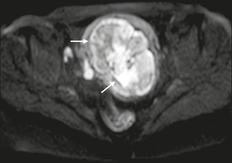
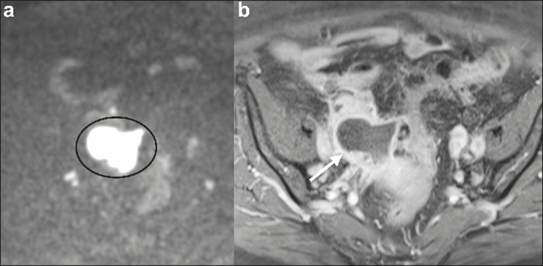
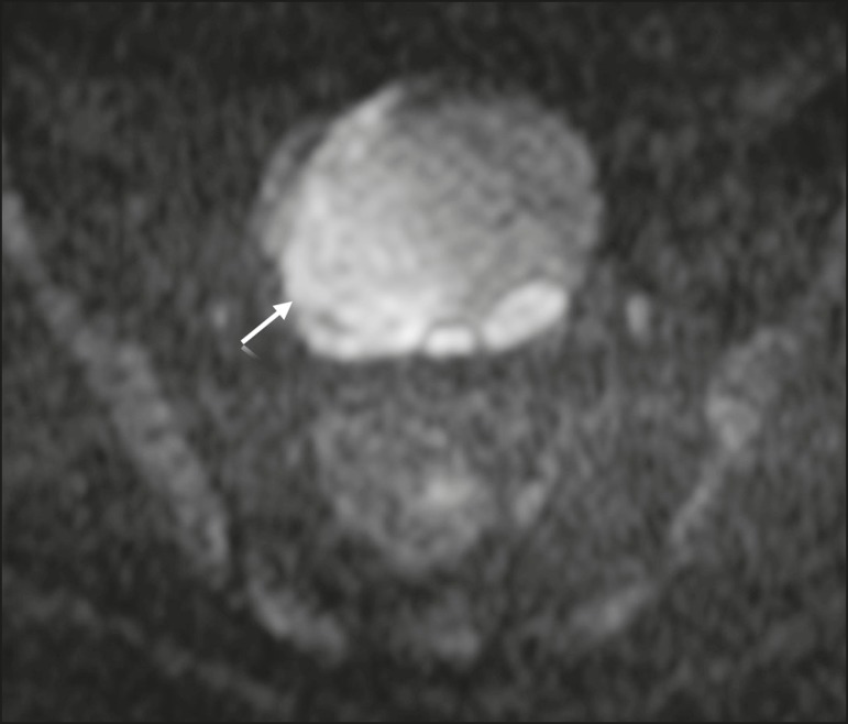
Diffusion-weighted imaging (DWI) is widely used in protocols for magnetic resonance imaging (MRI) of the female pelvis. It provides functional and structural information about biological tissues, without the use of ionizing radiation or intravenous administration of contrast medium. High signal intensity on DWI with simultaneous low signal intensity on apparent diffusion coefficient maps is usually associated with malignancy. However, that pattern can also be seen in many benign lesions, a fact that should be recognized by radiologists. Correlating DWI findings with those of conventional (T1- and T2-weighted) MRI sequences and those of contrast-enhanced MRI sequences is mandatory in order to avoid potential pitfalls. The aim of this review article is the description of the most relevant physiological and benign pathological conditions of the female pelvis that can show restricted diffusion on DWI.
Keywords: Diffusion magnetic resonance imaging; Magnetic resonance imaging; Pelvis/diagnostic imaging.
Figures


References
-
- Koh DM, Collins DJ. Diffusion-weighted MRI in the body: applications and challenges in oncology. AJR Am J Roentgenol. 2007;188:1622–1635. - PubMed
-
- Nougaret S, Tirumani SH, Addley H, et al. Pearls and pitfalls in MRI of gynecologic malignancy with diffusion-weighted technique. AJR Am J Roentgenol. 2013;200:261–276. - PubMed
-
- Thomassin-Naggara I, Fournier LS, Roussel A, et al. IRM de diffusion et pelvis féminin. J Radiol. 2010;91:431–440. - PubMed
-
- Feuerlein S, Pauls S, Juchems MS, et al. Pitfalls in abdominal diffusion-weighted imaging: how predictive is restricted water diffusion for malignancy. AJR Am J Roentgenol. 2009;193:1070–1076. - PubMed
Publication types
LinkOut - more resources
Full Text Sources
Other Literature Sources