. 2018 Mar 13:13:1495-1504.
doi: 10.2147/IJN.S157082. eCollection 2018.

The theranostic efficiency of tumor-specific, pH-responsive, peptide-modified, liposome-containing paclitaxel and superparamagnetic iron oxide nanoparticles

Affiliations

The theranostic efficiency of tumor-specific, pH-responsive, peptide-modified, liposome-containing paclitaxel and superparamagnetic iron oxide nanoparticles

Xiu-Chai Zheng et al. Int J Nanomedicine. .

Abstract

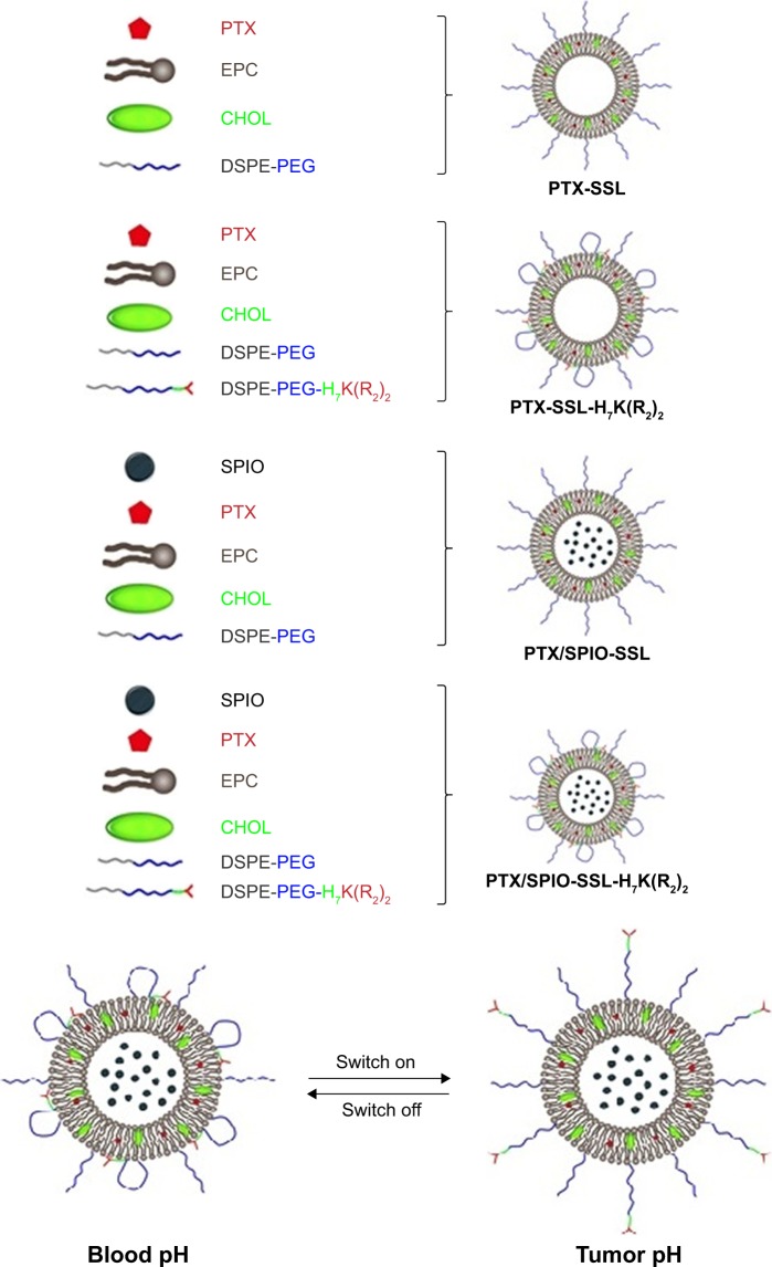
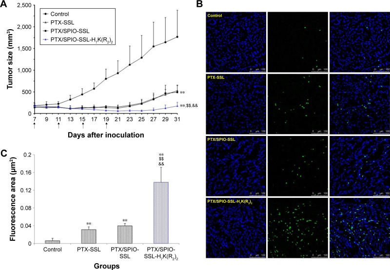
Background: In the present study, the tumor-specific, pH-responsive peptide H7K(R2)2-modified, theranostic liposome-containing paclitaxel (PTX) and superparamagnetic iron oxide nanoparticles (SPIO NPs), PTX/SPIO-SSL-H7K(R2)2, was prepared by using H7K(R2)2 as the targeting ligand, SPIO NPs as the magnetic resonance imaging (MRI) agent, PTX as antitumor drug.

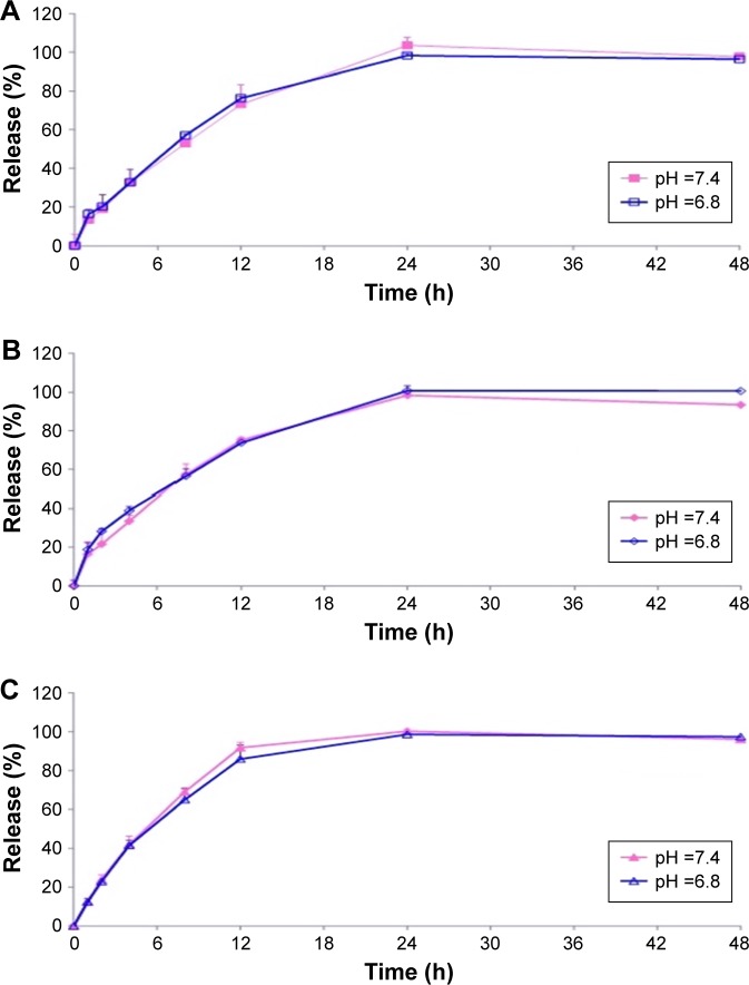
Methods: The PTX/SPIO-SSL-H7K(R2)2 was prepared by a thin film hydration method. The characteristics of PTX/SPIO-SSL-H7K(R2)2 were evaluated. The targeting effect, MRI, and antitumor activity of PTX/SPIO-SSL-H7K(R2)2 were investigated detail in vitro and in vivo in human breast carcinoma MDA-MB-231 cell models.

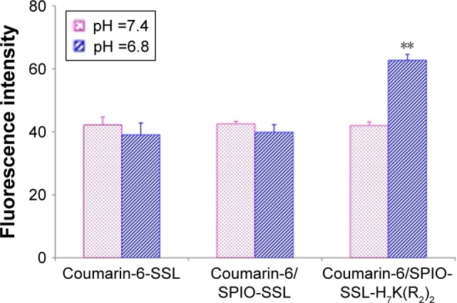
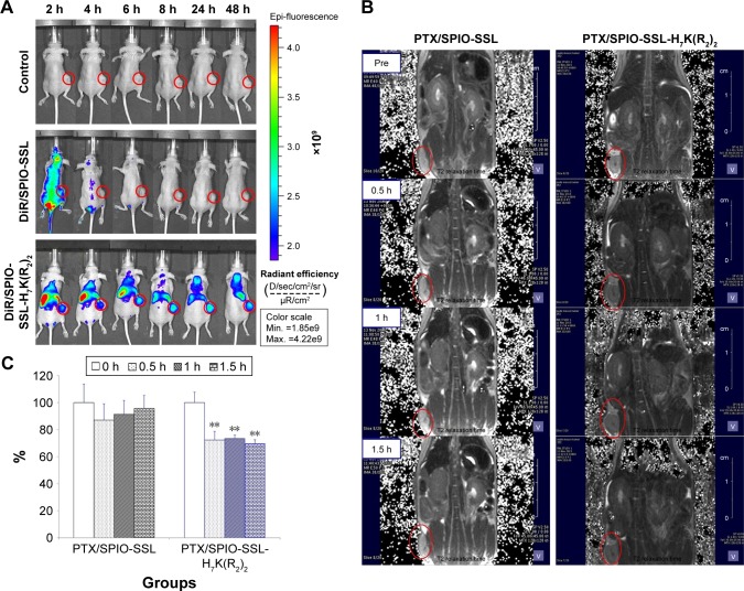
Results: Our results of in vitro flow cytometry, in vivo imaging, and in vivo MR imaging confirmed the pH-responsive characteristic of H7K(R2)2 in MDA-MB-231 cell line in vitro and in vivo. The results of in vivo MRI and in vivo antitumor activity confirmed the theranostic effect of PTX/SPIO-SSL-H7K(R2)2 in MDA-MB-231 tumor-bearing model.

Conclusion: Considering all our in vitro and in vivo results, we conclude that we developed targeting modified theranostic liposome which could achieve both role of antitumor and MRI.

Keywords: liposome; paclitaxel; superparamagnetic iron oxide nanoparticles; theranostic efficiency; tumor-specific pH-responsive peptide.


Conflict of interest statement

Disclosure The authors report no conflicts of interest in this work.

Figures

Figure 1
Figure 1
The schematic diagram for preparation of PTX-SSL, PTX-SSL-H7K(R2)2, PTX/SPIO-SSL, and PTX/SPIO-SSL-H7K(R2)2. Abbreviations: CHOL, cholesterol; EPC, egg phosphatidylcholine; PEG, polyethylene glycol; PTX, paclitaxel; PTX/SPIO-SSL, PTX/SPIO-loaded liposome; PTX/SPIO-SSL-H7K(R2)2, H7K(R2)2-modified liposome containing PTX and SPIO; PTX-SSL, sterically stabilized liposome containing PTX; SSL, sterically stabilized liposome; SPIO, superparamagnetic iron oxide.
Figure 2
Figure 2
In vitro release of PTX from PTX-SSL, PTX/SPIO-SSL, and PTX/SPIO-SSL-H7K(R2)2 at 37°C in pH 6.8 and 7.4 PBS buffer medium, respectively. Notes: Data represent the mean ± SD (n=3). (A) PTX-SSL; (B) PTX/SPIO-SSL; (C) PTX/SPIO-SSL-H7K(R2)2. Abbreviations: PTX, paclitaxel; PTX/SPIO-SSL, PTX/SPIO-loaded liposome; PTX/SPIO-SSL-H7K(R2)2, H7K(R2)2-modified liposome containing PTX and SPIO; PTX-SSL, sterically stabilized liposome containing PTX; SSL, sterically stabilized liposome; SPIO, superparamagnetic iron oxide.
Figure 3
Figure 3
The flow cytometric measurement. Notes: Flow cytometric measurement of coumarin-6 uptake from coumarin-6-SSL, coumarin-6/SPIO-SSL, or coumarin-6/SPIO-SSL-H7K(R2)2 by MDA-MB-231 cells (n=3). **p<0.01 versus that at pH 7.4. Abbreviations: SPIO, superparamagnetic iron oxide; SSL, sterically stabilized liposome.
Figure 4
Figure 4
Tissue distribution and in vivo MRI of magnetoliposomes in MDA-MB-231 tumor-bearing mice. Notes: (A) Tissue distribution of DiR-loaded liposomes in tumor-bearing mice. In vivo imaging of MDA-MB-231 tumor-bearing mice after 5% glucose solution (as control), DiR/SPIO-SSL, and DiR/SPIO-SSL-H7K(R2)2 administration at 2, 4, 6, 8, 24, and 48 h, respectively. (B) In vivo MRI of MDA-MB-231 tumor model. In vivo MRI of MDA-MB-231 tumor-bearing mice after PTX/SPIO-SSL and PTX/SPIO-SSL-H7K(R2)2 administration at 0, 0.5, 1, and 1.5 h, respectively. (C) Quantitative T2 measurements in in vivo MRI of MDA-MB-231 tumor model. Quantitative T2 measurements showed ~30%±3% decrease of PTX/SPIO-SSL-H7K(R2)2 group at 0.5, 1, and 1.5 h. **p<0.01 versus that of preinjection of liposomes (0 h time point) as control. Abbreviations: DiR, 1,10-dioctadecyltetramethyl indotricarbocyanine iodide; MRI, magnetic resonance imaging; PTX, paclitaxel; PTX/SPIO-SSL, PTX/SPIO-loaded liposome; PTX/SPIO-SSL-H7K(R2)2, H7K(R2)2-modified liposome containing PTX and SPIO; SPIO, superparamagnetic iron oxide; SSL, sterically stabilized liposome.
Figure 5
Figure 5
In vivo antitumor activity of PTX/SPIO-SSL-H7K(R2)2. Notes: BALB/C nude mice were inoculated s.c. with MDA-MB-231 cells and treated with 5% glucose solution, PTX-SSL (15 mg/kg, i.v., q4d), PTX/SPIO-SSL (15 mg/kg, i.v., q4d), and PTX/SPIO-SSL-H7K(R2)2 (15 mg/kg, i.v., q4d), respectively. For each administration, formulations were given to mice via the tail vein. (A) Tumor growth inhibition. (B) TUNEL staining of the tumor tissue sections was performed according to the standard protocols provided by the manufacturers. The apoptotic cells were detected by TUNEL. DNA strand breaks were labeled green, and the nuclei were stained with Hoechst 332589 (blue). Apoptotic cells exhibited a turquoise color as a result of color merging of these two labels. (C) The fluorescence area of each group was used for the statistical analysis of apoptosis activity. **p<0.01 versus the 5% glucose solution treatment group as a control; $$p<0.01 versus the PTX-SSL treatment group; &&p<0.01 versus the PTX/SPIO-SSL treatment group. Abbreviations: i.v., intravenous; PTX, paclitaxel; PTX/SPIO-SSL, PTX/SPIO-loaded liposome; PTX/SPIO-SSL-H7K(R2)2, H7K(R2)2-modified liposome containing PTX and SPIO; PTX-SSL, sterically stabilized liposome containing PTX; q4d, twice a day for 4 days; s.c., subcutaneously; SPIO, superparamagnetic iron oxide; TUNEL, terminal deoxynucleotidyl transferase dUTP nick end labeling.

References

    1. Ryu JH, Koo H, Sun IC, et al. Tumor-targeting multi-functional nanoparticles for theragnosis: new paradigm for cancer therapy. Adv Drug Deliv Rev. 2012;64(13):1447–1458. - PubMed
    1. Schleich N, Danhier F, Préat V. Iron oxide-loaded nanotheranostics: major obstacles to in vivo studies and clinical translation. J Control Release. 2015;198:35–54. - PubMed
    1. Hahn MA, Singh AK, Sharma P, Brown SC, Moudgil BM. Nanoparticles as contrast agents for in-vivo bioimaging: current status and future perspectives. Anal Bioanal Chem. 2011;399(1):3–27. - PubMed
    1. Bennett KM, Jo J, Cabral H, Bakalova R, Aoki I. MR imaging techniques for nano-pathophysiology and theranostics. Adv Drug Deliv Rev. 2014;74:75–94. - PubMed
    1. Roy Chowdhury M, Schumann C, Bhakta-Guha D, Guha G. Cancer nanotheranostics: strategies, promises and impediments. Biomed Pharmacother. 2016;84:291–304. - PubMed

MeSH terms

LinkOut - more resources