

Neural Networks Mediating High-Level Mentalizing in Patients With Right Cerebral Hemispheric Gliomas
- PMID: 29559899
- PMCID: PMC5845682
- DOI: 10.3389/fnbeh.2018.00033
Neural Networks Mediating High-Level Mentalizing in Patients With Right Cerebral Hemispheric Gliomas
Abstract
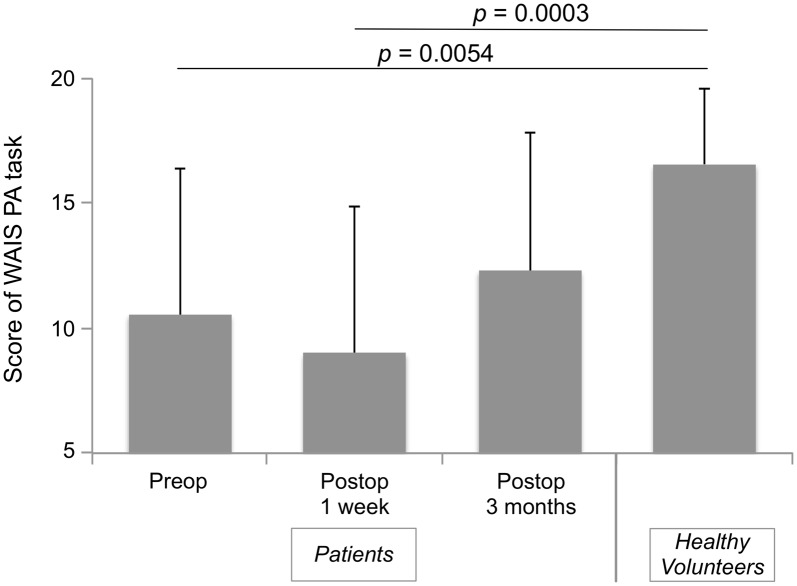
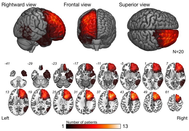
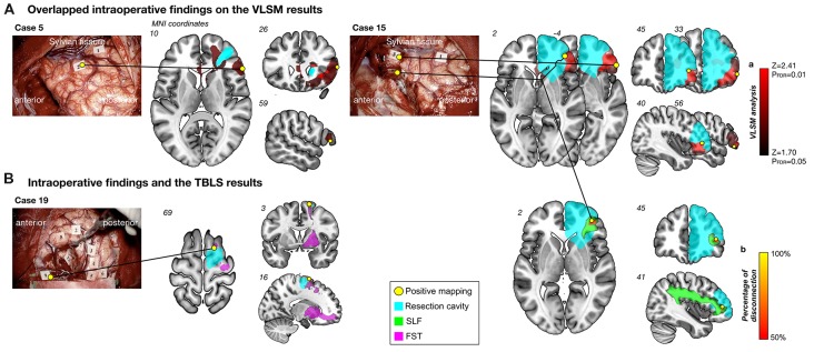
Mentalizing is the ability to understand others' mental state through external cues. It consists of two networks, namely low-level and high-level metalizing. Although it is an essential function in our daily social life, surgical resection of right cerebral hemisphere disturbs mentalizing processing with high possibility. In the past, little was known about the white matter related to high-level mentalizing, and the conservation of high-level mentalizing during surgery has not been a focus of attention. Therefore, the main purpose of this study was to examine the neural networks underlying high-level mentalizing and then, secondarily, investigate the usefulness of awake surgery in preserving the mentalizing network. A total of 20 patients with glioma localized in the right hemisphere who underwent awake surgery participated in this study. All patients were assigned to two groups: with or without intraoperative assessment of high-level mentalizing. Their high-level mentalizing abilities were assessed before surgery and 1 week and 3 months after surgery. At 3 months after surgery, only patients who received the intraoperative high-level mentalizing test showed the same score as normal healthy volunteers. The tract-based lesion symptom analysis was performed to confirm the severity of damage of associated fibers and high-level mentalizing accuracy. This analysis revealed the superior longitudinal fascicles (SLF) III and fronto-striatal tract (FST) to be associated with high-level mentalizing processing. Moreover, the voxel-based lesion symptom analysis demonstrated that resection of orbito-frontal cortex (OFC) causes persistent mentalizing dysfunction. Our study indicates that damage of the OFC and structural connectivity of the SLF and FST causes the disorder of mentalizing after surgery, and assessing high-level mentalizing during surgery may be useful to preserve these pathways.
Keywords: awake surgery; fronto-striatal tract; glioma; mentalizing; superior longitudinal fascicle.
Figures

Similar articles
-
Neuropsychological evidence for the crucial role of the right arcuate fasciculus in the face-based mentalizing network: A disconnection analysis.Neuropsychologia. 2018 Jul 1;115:179-187. doi: 10.1016/j.neuropsychologia.2018.01.024. Epub 2018 Jan 31. Neuropsychologia. 2018. PMID: 29360518
-
Chronic spatial working memory deficit associated with the superior longitudinal fasciculus: a study using voxel-based lesion-symptom mapping and intraoperative direct stimulation in right prefrontal glioma surgery.J Neurosurg. 2016 Oct;125(4):1024-1032. doi: 10.3171/2015.10.JNS1591. Epub 2016 Feb 19. J Neurosurg. 2016. PMID: 26894458
-
Neural pathways subserving face-based mentalizing.Brain Struct Funct. 2017 Sep;222(7):3087-3105. doi: 10.1007/s00429-017-1388-0. Epub 2017 Feb 27. Brain Struct Funct. 2017. PMID: 28243761
-
Critical Neural Networks in Awake Surgery for Gliomas.Neurol Med Chir (Tokyo). 2016 Nov 15;56(11):674-686. doi: 10.2176/nmc.ra.2016-0069. Epub 2016 Jun 2. Neurol Med Chir (Tokyo). 2016. PMID: 27250817 Free PMC article. Review.
-
Challenging the Myth of Right Nondominant Hemisphere: Lessons from Corticosubcortical Stimulation Mapping in Awake Surgery and Surgical Implications.World Neurosurg. 2017 Jul;103:449-456. doi: 10.1016/j.wneu.2017.04.021. Epub 2017 Apr 15. World Neurosurg. 2017. PMID: 28419879 Review.
Cited by
-
The superior longitudinal fascicle: reconsidering the fronto-parietal neural network based on anatomy and function.Brain Imaging Behav. 2020 Dec;14(6):2817-2830. doi: 10.1007/s11682-019-00187-4. Brain Imaging Behav. 2020. PMID: 31468374
-
Glioblastomas at the white matter of temporo-parietal junction cause a poor postoperative independence level.J Neurooncol. 2023 Oct;165(1):191-199. doi: 10.1007/s11060-023-04479-0. Epub 2023 Oct 17. J Neurooncol. 2023. PMID: 37847481
-
Sociocognitive Functioning and Psychosocial Burden in Patients with Brain Tumors.Cancers (Basel). 2022 Feb 1;14(3):767. doi: 10.3390/cancers14030767. Cancers (Basel). 2022. PMID: 35159034 Free PMC article. Review.
-
An update on tests used for intraoperative monitoring of cognition during awake craniotomy.Acta Neurochir (Wien). 2024 May 7;166(1):204. doi: 10.1007/s00701-024-06062-6. Acta Neurochir (Wien). 2024. PMID: 38713405 Free PMC article.
-
Role of Preoperative Assessment in Predicting Tumor-Induced Plasticity in Patients with Diffuse Gliomas.J Clin Med. 2021 Mar 7;10(5):1108. doi: 10.3390/jcm10051108. J Clin Med. 2021. PMID: 33799925 Free PMC article.
References
-
- Apperly I. (2012). Mindreaders: The Cognitive Basis of “Theory of Mind”. New York, NY: Psychology Press.
LinkOut - more resources
Full Text Sources
Other Literature Sources
Miscellaneous