

Case Reports
doi: 10.1155/2018/6068258.
eCollection 2018.
Tuberculous Enteritis Presenting as Acute Appendicitis and Perirectal Abscess
Affiliations
- PMID: 29560012
- PMCID: PMC5845528
- DOI: 10.1155/2018/6068258
Item in Clipboard
Case Reports
Tuberculous Enteritis Presenting as Acute Appendicitis and Perirectal Abscess
Case Rep Med.
.
Abstract
Mycobacterium tuberculosis has a wide variety of presentations. A rare occurrence is gastrointestinal tuberculosis. It may occur anywhere along the alimentary canal but usually occurs in the ileocecum with rare involvement of the appendix.
Figures
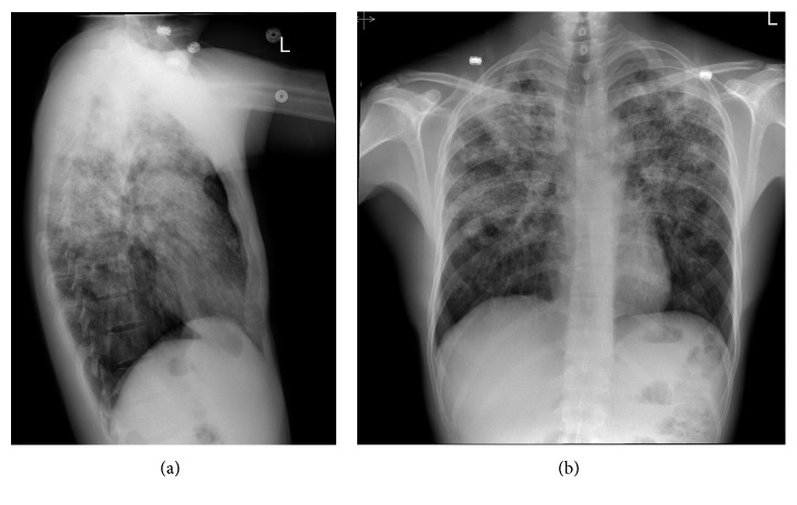
(a, b) PA CXR showing biapical pleural thickening associated with parenchymal scarring, bronchiectasis, nodularity, and superimposed infiltrates.
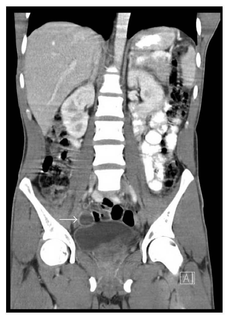
Solid white arrow outlining fluid-filled appendix suggestive of appendicitis.
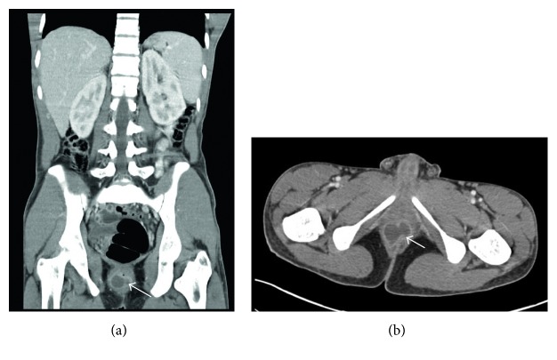
Solid dense arrow delineating perirectal abscess in the saggital (a) and coronal (b) view of the abdominal CT scan.
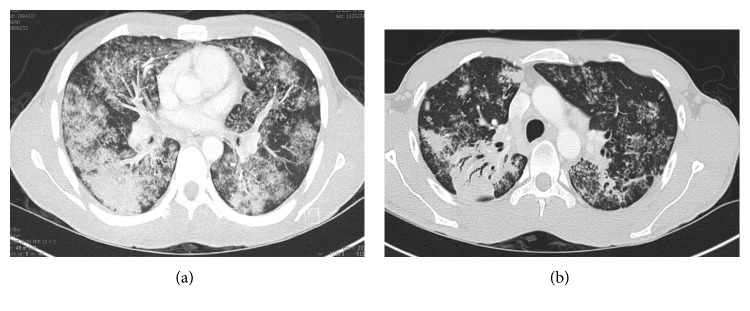
(a) Diffuse airspace patchy consolidation with distention of bronchi and fluffy infiltrates. (b) Dense consolidation in the superior aspect of the right lower lobe with air bronchograms.
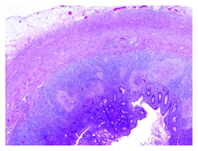
Granulomatous appendix (2x; 20 magnification).
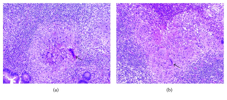
(a, b) H&E stain showing granulomatous inflammation of the appendix with solid black arrows depicting multinucleated giant cells. Central necrosis can be seen in image A outlined by solid white arrow (10x; 100).
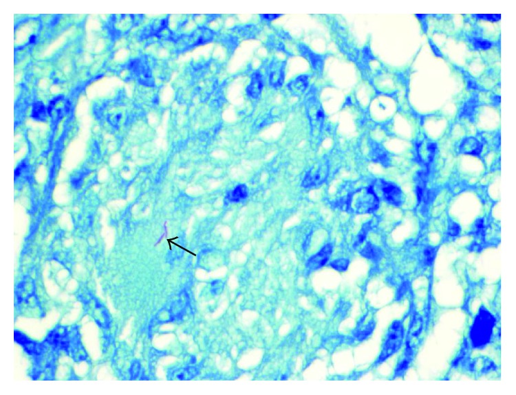
Solid black arrow depicting Ziehl-Neelsen AFB stain positive for acid-fast positive bacilli in the cytoplasm of a multinucleated giant cell (60x; 600).
Similar articles
-
Tuberculous appendicitis presenting with lower gastrointestinal hemorrhage--a case report and review of the literature.J Med Assoc Thai. 2008 Jun;91(6):937-42. J Med Assoc Thai. 2008. PMID: 18697397 Review.
-
Tuberculous appendix: a review of clinical presentations and outcomes.Singapore Med J. 2011 Feb;52(2):90-3. Singapore Med J. 2011. PMID: 21373734
-
Tuberculous appendicitis. A case report.Med Ultrason. 2017 May 3;19(3):333-335. doi: 10.11152/mu-1085. Med Ultrason. 2017. PMID: 28845503
-
[Acute perforated appendicitis secondary to tuberculosis as a cause of postpartum infection].Rev Chilena Infectol. 2020 Apr;37(2):186-189. doi: 10.4067/s0716-10182020000200186. Rev Chilena Infectol. 2020. PMID: 32730488 Spanish.
-
Isolated tuberculous liver abscess in an immunocompetent adult patient: A case report and literature review.J Microbiol Immunol Infect. 2016 Jun;49(3):455-8. doi: 10.1016/j.jmii.2013.09.003. Epub 2013 Nov 11. J Microbiol Immunol Infect. 2016. PMID: 24231587 Review.
Cited by
-
Diagnostic Accuracy of Abdominal Ultrasonography in Pediatric Acute Appendicitis.Bull Emerg Trauma. 2019 Jul;7(3):278-283. doi: 10.29252/beat-0703011. Bull Emerg Trauma. 2019. PMID: 31392228 Free PMC article.
-
An atlas overview of characteristic features of tuberculosis that may be encountered at autopsy.Forensic Sci Med Pathol. 2020 Mar;16(1):143-151. doi: 10.1007/s12024-019-00161-y. Epub 2019 Aug 30. Forensic Sci Med Pathol. 2020. PMID: 31471869 Review.
-
Tuberculous appendicitis: A review of reported cases over the past 10 years.J Clin Tuberc Other Mycobact Dis. 2021 Mar 18;23:100228. doi: 10.1016/j.jctube.2021.100228. eCollection 2021 May. J Clin Tuberc Other Mycobact Dis. 2021. PMID: 33898762 Free PMC article. Review.
References
Publication types
LinkOut - more resources
Full Text Sources
Other Literature Sources
