

Paeoniflorin exerts neuroprotective effects by modulating the M1/M2 subset polarization of microglia/macrophages in the hippocampal CA1 region of vascular dementia rats via cannabinoid receptor 2
- PMID: 29560022
- PMCID: PMC5859430
- DOI: 10.1186/s13020-018-0173-1
Paeoniflorin exerts neuroprotective effects by modulating the M1/M2 subset polarization of microglia/macrophages in the hippocampal CA1 region of vascular dementia rats via cannabinoid receptor 2
Abstract
Background: Cerebral hypoperfusion is a pivotal risk factor for vascular dementia (VD), for which effective therapy remains inadequate. Persistent inflammatory responses and excessive chemotaxis of microglia/macrophages in the brain may accelerate the progression of VD. Endocannabinoids are involved in neuronal protection against inflammation-induced neuronal injury. Cannabinoids acting at cannabinoid receptor 2 (CB2R) can decrease inflammation. Based on the identification of paeoniflorin (PF) as a CB2R agonist, we investigated the neuroprotective and microglia/macrophages M1 to M2 polarization promoting effects of PF in a permanent four-vessel occlusion rat model.
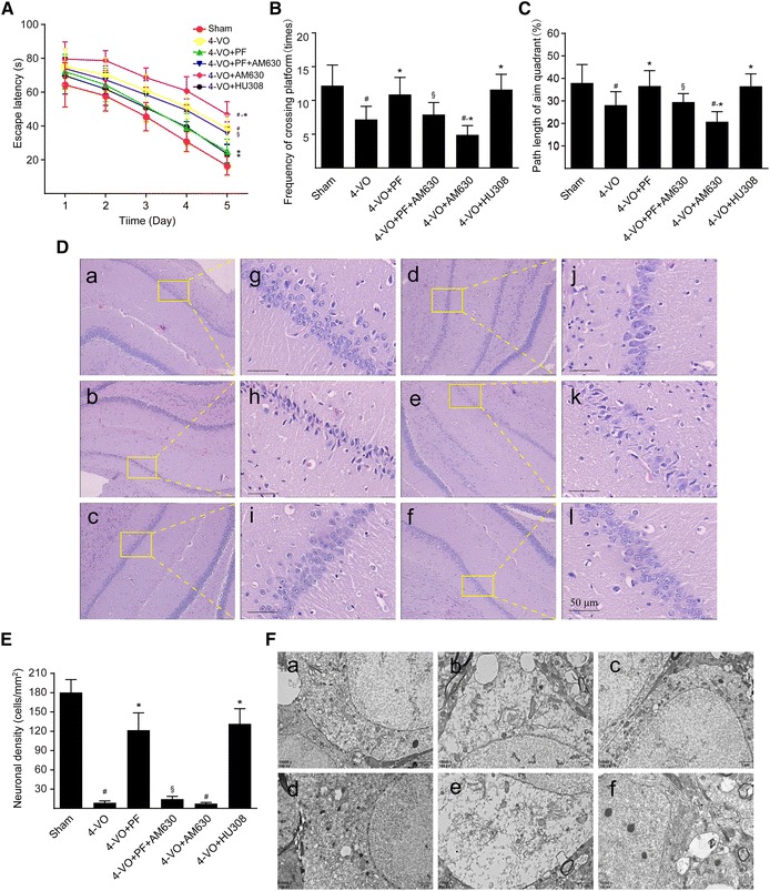
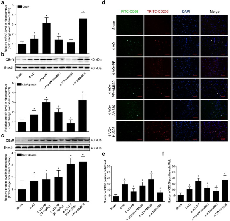
Methods: One week after surgery, PF was intraperitoneally administered at a dose of 40 mg/kg once a day for 28 successive days. The effects of PF on memory deficit were investigated by a Morris water maze test, and the effects of PF on hippocampal neuronal damage were evaluated by light microscope and electron microscope. The mRNA and protein expression levels of key molecules related to the M1/M2 polarization of microglia/macrophages were assessed by RT-qPCR and Western blotting, respectively.
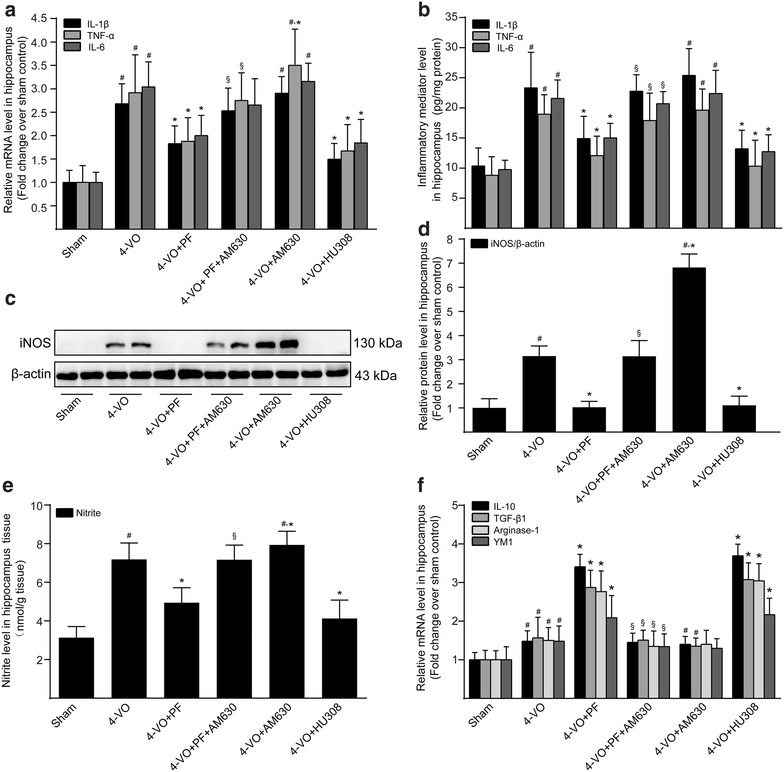
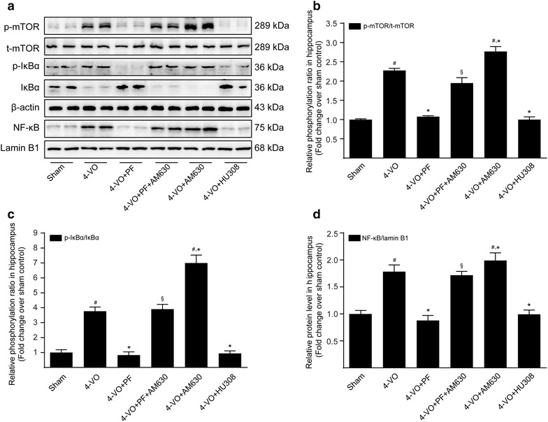
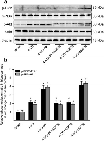
Results: Administration of PF could significantly attenuate cerebral hypoperfusion-induced impairment of learning and memory and reduce the morphological and ultrastructural changes in the hippocampal CA1 region of rats. Moreover, PF promoted an M1 to M2 phenotype transition in microglia/macrophages in the hippocampus of rats. In addition to its inhibitory property against proinflammatory M1 mediator expression, such as IL-1β, IL-6, TNF-α and NO, PF dramatically up-regulated expression of anti-inflammatory cytokines IL-10 and TGF-β1. Importantly, CB2R antagonist AM630 abolished these beneficial effects produced by PF on learning, memory and hippocampus structure in rats, as well as the polarization of microglia/macrophages to the M2 phenotype. Additionally, PF treatment significantly inhibited cerebral hypoperfusion-induced mTOR/NF-κB proinflammatory pathway and enhanced PI3K/Akt anti-inflammatory pathway. Effects of PF on these signaling pathways were effectively attenuated when rats were co-treated with PF and AM630, indicating that the mTOR/NF-κB and PI3K/Akt signaling pathways were involved in the PF effects through CB2R activation.
Conclusion: These findings demonstrated PF exerts its neuroprotective effect and shifts the inflammatory milieu toward resolution by modulation of microglia/macrophage polarization via CB2R activation.
Keywords: Cannabinoid receptor 2; Neuroprotection; Paeoniflorin; Vascular dementia.
Figures
Similar articles
-
Cysteinyl Leukotriene Receptor 2 is Involved in Inflammation and Neuronal Damage by Mediating Microglia M1/M2 Polarization through NF-κB Pathway.Neuroscience. 2019 Dec 1;422:99-118. doi: 10.1016/j.neuroscience.2019.10.048. Epub 2019 Nov 11. Neuroscience. 2019. PMID: 31726033
-
Paeoniflorin improves regional cerebral blood flow and suppresses inflammatory factors in the hippocampus of rats with vascular dementia.Chin J Integr Med. 2017 Sep;23(9):696-702. doi: 10.1007/s11655-015-2124-3. Epub 2015 Nov 17. Chin J Integr Med. 2017. PMID: 26577108
-
Cannabinoid receptor-2 stimulation suppresses neuroinflammation by regulating microglial M1/M2 polarization through the cAMP/PKA pathway in an experimental GMH rat model.Brain Behav Immun. 2016 Nov;58:118-129. doi: 10.1016/j.bbi.2016.05.020. Epub 2016 May 31. Brain Behav Immun. 2016. PMID: 27261088
-
Exploring the potential of treating chronic liver disease targeting the PI3K/Akt pathway and polarization mechanism of macrophages.Heliyon. 2023 Jun 9;9(6):e17116. doi: 10.1016/j.heliyon.2023.e17116. eCollection 2023 Jun. Heliyon. 2023. PMID: 37484431 Free PMC article. Review.
-
Manipulating Macrophage/Microglia Polarization to Treat Glioblastoma or Multiple Sclerosis.Pharmaceutics. 2022 Feb 1;14(2):344. doi: 10.3390/pharmaceutics14020344. Pharmaceutics. 2022. PMID: 35214076 Free PMC article. Review.
Cited by
-
A Novel Trichinella spiralis Galectin Strengthens the Macrophage ADCC Killing of Larvae via Driving M1 Polarization.Int J Mol Sci. 2024 Oct 10;25(20):10920. doi: 10.3390/ijms252010920. Int J Mol Sci. 2024. PMID: 39456703 Free PMC article.
-
CB2R agonist GW405833 alleviates acute liver failure in mice via inhibiting HIF-1α-mediated reprogramming of glycometabolism and macrophage proliferation.Acta Pharmacol Sin. 2023 Jul;44(7):1391-1403. doi: 10.1038/s41401-022-01037-8. Epub 2023 Jan 25. Acta Pharmacol Sin. 2023. PMID: 36697976 Free PMC article.
-
Naomaitai Ameliorated Brain Damage in Rats with Vascular Dementia by PI3K/PDK1/AKT Signaling Pathway.Evid Based Complement Alternat Med. 2019 Feb 5;2019:2702068. doi: 10.1155/2019/2702068. eCollection 2019. Evid Based Complement Alternat Med. 2019. PMID: 30867669 Free PMC article.
-
Rubus fruticosus leaf extract inhibits vascular dementia-induced memory impairment and neuronal loss by attenuating neuroinflammation.Anat Cell Biol. 2023 Dec 31;56(4):494-507. doi: 10.5115/acb.23.195. Epub 2023 Sep 25. Anat Cell Biol. 2023. PMID: 37743615 Free PMC article.
-
The Role of Oxidative Stress and Inflammation in the Pathogenesis and Treatment of Vascular Dementia.Cells. 2025 Apr 17;14(8):609. doi: 10.3390/cells14080609. Cells. 2025. PMID: 40277934 Free PMC article. Review.
References
-
- Black SE. Vascular cognitive impairment: epidemiology, subtypes, diagnosis and management. J R Coll Surg Edinb. 2011;41:49–56. - PubMed
LinkOut - more resources
Full Text Sources
Other Literature Sources
Miscellaneous
