

Reconstitution of high-grade serous ovarian carcinoma from primary fallopian tube secretory epithelial cells
- PMID: 29560094
- PMCID: PMC5849158
- DOI: 10.18632/oncotarget.23035
Reconstitution of high-grade serous ovarian carcinoma from primary fallopian tube secretory epithelial cells
Abstract
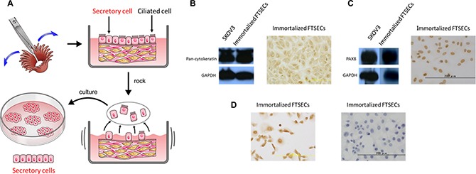
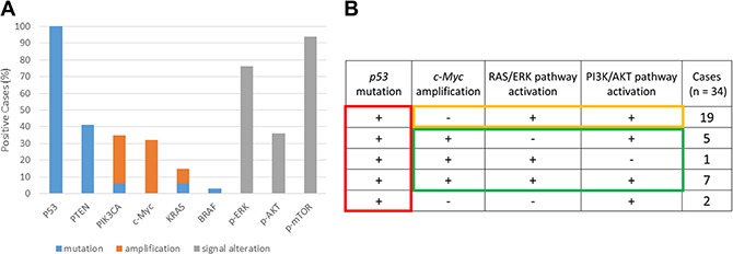
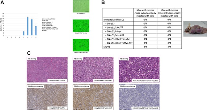
Fallopian tube secretory epithelial cells (FTSECs) have been suggested to be the source of high-grade serous ovarian carcinoma (HGSOC). Although several genetic alterations are known to be involved in HGSOC development, the minimal requirements remain unclear. We aimed to identify oncogenic mutations indispensable for HGSOC development in a stepwise model, using immortalized FTSECs. FTSECs were isolated from clinical samples and immortalized by overexpression of cyclin D1, CDK4R24C, and hTERT. Oncogenic mutations in the p53, c-Myc, and RAS/PI3K pathways were mimicked by lentiviral transduction. We found two distinct patterns of gene alteration essential for HGSOC development: p53/KRAS/AKT and p53/KRAS/c-Myc. Dominant-negative p53, alone or combined with oncogenic KRAS (KRASV12), constitutively active AKT (CA-AKT), and c-Myc, did not induce tumorigenesis in immortalized cells; however, overexpression of CA-AKT or c-Myc, along with dominant-negative p53 and KRASV12, conferred tumorigenic potential. Transformed FTSECs formed tumors in nude mice that were grossly, histologically, and immunohistochemically similar to human HGSOCs. Interestingly, mice harboring tumors with c-Myc amplifications displayed extensive metastases, consistent with the increased dissemination in their human counterparts. Thus, aberrant p53/KRASV12/c-Myc or p53/KRASV12/PI3K-AKT signaling was the minimum requirement for FTSEC carcinogenesis. The model based on this evidence could shed light on the early stages of HGSOC development.
Keywords: RAS/PI3K pathway; c-Myc; carcinogenesis; fallopian tube; high-grade serous ovarian carcinoma.
Conflict of interest statement
CONFLICTS OF INTEREST None.
Figures

Similar articles
-
Modeling high-grade serous ovarian carcinogenesis from the fallopian tube.Proc Natl Acad Sci U S A. 2011 May 3;108(18):7547-52. doi: 10.1073/pnas.1017300108. Epub 2011 Apr 18. Proc Natl Acad Sci U S A. 2011. PMID: 21502498 Free PMC article.
-
Chronic iron exposure and c-Myc/H-ras-mediated transformation in fallopian tube cells alter the expression of EVI1, amplified at 3q26.2 in ovarian cancer.Oncogenesis. 2019 Aug 21;8(9):46. doi: 10.1038/s41389-019-0154-y. Oncogenesis. 2019. PMID: 31434871 Free PMC article.
-
MYC is sufficient to generate mid-life high-grade serous ovarian and uterine serous carcinomas in a p53-R270H mouse model.bioRxiv [Preprint]. 2024 Jan 29:2024.01.24.576924. doi: 10.1101/2024.01.24.576924. bioRxiv. 2024. Update in: Cancer Res Commun. 2024 Sep 1;4(9):2525-2538. doi: 10.1158/2767-9764.CRC-24-0144. PMID: 38352443 Free PMC article. Updated. Preprint.
-
Studying platinum sensitivity and resistance in high-grade serous ovarian cancer: Different models for different questions.Drug Resist Updat. 2016 Jan;24:55-69. doi: 10.1016/j.drup.2015.11.005. Epub 2015 Nov 26. Drug Resist Updat. 2016. PMID: 26830315 Review.
-
Fallopian tube precursors of ovarian low- and high-grade serous neoplasms.Histopathology. 2013 Jan;62(1):44-58. doi: 10.1111/his.12046. Histopathology. 2013. PMID: 23240669 Review.
Cited by
-
Systematic Identification of Characteristic Genes of Ovarian Clear Cell Carcinoma Compared with High-Grade Serous Carcinoma Based on RNA-Sequencing.Int J Mol Sci. 2019 Sep 4;20(18):4330. doi: 10.3390/ijms20184330. Int J Mol Sci. 2019. PMID: 31487856 Free PMC article.
-
TP53 loss initiates chromosomal instability in fallopian tube epithelial cells.Dis Model Mech. 2021 Nov 1;14(11):dmm049001. doi: 10.1242/dmm.049001. Epub 2021 Nov 30. Dis Model Mech. 2021. PMID: 34569598 Free PMC article.
-
Establishment of five immortalized human ovarian surface epithelial cell lines via SV40 T antigen or HPV E6/E7 expression.PLoS One. 2018 Oct 8;13(10):e0205297. doi: 10.1371/journal.pone.0205297. eCollection 2018. PLoS One. 2018. PMID: 30296284 Free PMC article.
-
Aberrant SKP1 Expression: Diverse Mechanisms Impacting Genome and Chromosome Stability.Front Cell Dev Biol. 2022 Mar 8;10:859582. doi: 10.3389/fcell.2022.859582. eCollection 2022. Front Cell Dev Biol. 2022. PMID: 35345853 Free PMC article. Review.
-
Development of Low-Grade Serous Ovarian Carcinoma from Benign Ovarian Serous Cystadenoma Cells.Cancers (Basel). 2022 Mar 15;14(6):1506. doi: 10.3390/cancers14061506. Cancers (Basel). 2022. PMID: 35326657 Free PMC article.
References
-
- Wingo PA, Tong T, Bolden S. Cancer statistics 1995. CA: A CA Cancer J Clin. 1995;45:8–30. - PubMed
-
- Przybycin CG, Kurman RJ, Ronnett BM, Shih Ie M, Vang R. Are all pelvic (nonuterine) serous carcinomas of tubal origin? Am J Surg Pathol. 2010;34:1407–1416. - PubMed
-
- Kindelberger DW, Lee Y, Miron A, Hirsch MS, Feltmate C, Medeiros F, Callahan MJ, Garner EO, Gordon RW, Birch C, Berkowitz RS, Muto MG, Crum CP. Intraepithelial carcinoma of the fimbria and pelvic serous carcinoma: Evidence for a causal relationship. Am J Surg Pathol. 2007;31:161–169. - PubMed
-
- Tang S, Onuma K, Deb P, Wang E, Lytwyn A, Sur M, Daya D. Frequency of serous tubal intraepithelial carcinoma in various gynecologic malignancies: A study of 300 consecutive cases. Int J Gynecol Pathol. 2012;31:103–110. - PubMed
LinkOut - more resources
Full Text Sources
Other Literature Sources
Research Materials
Miscellaneous
