. 2018 Jan 3;9(16):12649-12661.
doi: 10.18632/oncotarget.23898. eCollection 2018 Feb 27.

Fucoidan-coated CuS nanoparticles for chemo-and photothermal therapy against cancer

Affiliations

Fucoidan-coated CuS nanoparticles for chemo-and photothermal therapy against cancer

Bian Jang et al. Oncotarget. .

Abstract

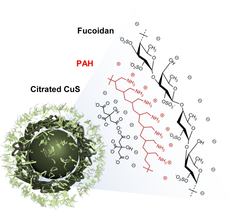
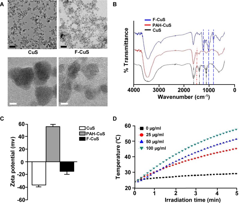
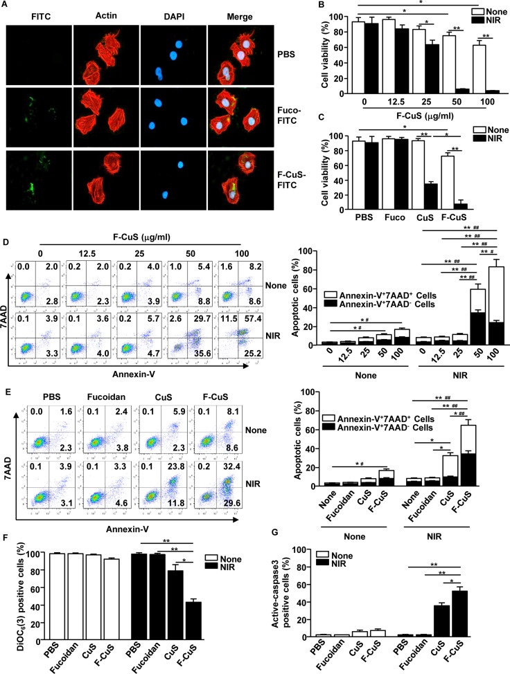
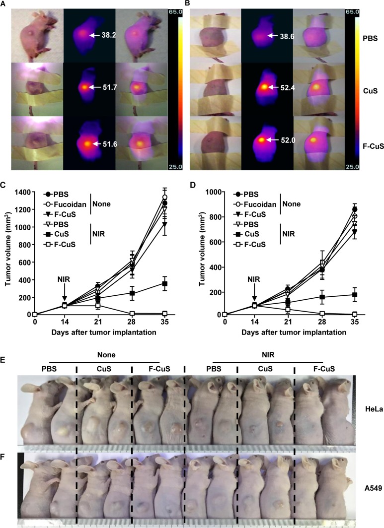
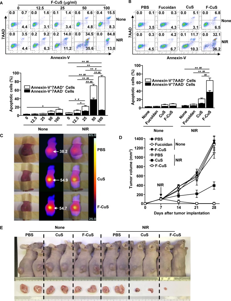
In advanced cancer therapy, the combinational therapeutic effect of photothermal therapy (PTT) using near-infrared (NIR) light-responsive nanoparticles (NPs) and anti-cancer drug delivery-mediated chemotherapy has been widely applied. In the present study, using a facile, low-cost, and solution-based method, we developed and synthesized fucoidan, a natural polymer isolated from seaweed that has demonstrated anti-cancer effect, and coated NPs with it as an ideal candidate in chemo-photothermal therapy against cancer cells. Fucoidan-coated copper sulfide nanoparticles (F-CuS) act not only as a nanocarrier to enhance the intracellular delivery of fucoidan but also as a photothermal agent to effectively ablate different cancer cells (e.g., HeLa, A549, and K562), both in vitro and in vivo, with the induction of apoptosis under 808 nm diode laser irradiation. These results point to the potential usage of F-CuS in treating human cancer.

Keywords: apoptosis; chemotherapy; copper sulfide nanoparticles; fucodian; photothermal therapy.


Conflict of interest statement

CONFLICTS OF INTEREST Competing financial interests: The authors declare no competing financial interests.

Figures

Figure 1
Figure 1. Structural illustration of F-CuS prepared for chemo- and photothermal cancer therapy
Figure 2
Figure 2. Characterization of F-CuS
(A) FE-TEM images of CuS and F-CuS. Scale bars: 50 nm (black) and 5 nm (white). (B) FT-IR spectra of CuS (black), PAH-CuS (red), and F-CuS (blue). (C) Zeta potential value of each NP. (D) Photothermal heating curves of different concentrations of F-CuS dispersed in water, irradiated for 5 min with an 808 nm laser at a power density of 2 W/cm2.
Figure 3
Figure 3. F-CuS-induced apoptosis of HeLa cells
(A) HeLa cells (2 × 10⁵) were incubated with fucoidan-FITC and F-CuS-FITC for 2 h. The cells were then stained with β-actin and 4′,6-Diamidino-2-Phenylindole, Dihydrochloride (DAPI). The FITC expression levels were measured with a confocal microscope. (B) HeLa cells were treated with an indicated concentration of F-CuS, and the cells were irradiated for 5 min with an 808 nm laser. Cell viability was analyzed by MTT assay. (C) MTT analysis of fucoidan (75 ng/mL), CuS (200 μg/mL), and F-CuS (50 μg/mL) -treated HeLa cells with or without NIR irradiation. (D) Dose-dependent apoptotic effect of F-CuS with or without NIR irradiation was analyzed by annexin-V and 7AAD staining (left panel). Mean percentages of early-apoptotic cells (annexin-V+7AAD cells) and late-apoptotic/necrotic cells (annexin-V+7AAD+ cells) are shown (right panel). #p < 0.05, ##p < 0.01 for early-apoptotic cells; *p < 0.05, **p < 0.01 for late-apoptotic/necrotic cells. (E) Apoptosis of HeLa cells following treatment with fucoidan, CuS, and F-CuS with or without NIR irradiation was analyzed through flow cytometry. The mean percentages of early-apoptotic cells (annexin-V+7AAD cells) and late-apoptotic/necrotic cells (annexin-V+7AAD+ cells) are shown (right panel). #p < 0.05, ##p < 0.01 for early-apoptotic cells; *p < 0.05, **p < 0.01 for late-apoptotic/necrotic cells. (F) Mitochondria permeability was measured by DiOC6(3) reduction in flow cytometry; *p < 0.05, **p < 0.01. (G) Activation of caspase-3 was analyzed by flow cytometry, and the mean percentages of positive cells are shown; *p < 0.05, **p < 0.01. All data are representative of, or the average of, analyses of six independent samples (i.e., two samples per experiment, three independent experiments).
Figure 4
Figure 4. Chemo–photothermal therapy by F-CuS
Nude mice were injected s.c. with 5 × 10⁶ HeLa cells and 5 × 10⁶ A549 cells. Once tumors were measured to be ~5.0 mm (after 14 d), the mice were treated i.t. with 4 μg/kg fucoidan, 10 mg/kg CuS, or 2.5 mg/kg F-CuS. Two hours after treatment, the mice were irradiated for 5 min with an 808 nm laser at 2 W/cm2. (A and B) Thermal image of HeLa tumor mice (A) and A549 tumor mice (B) are shown after NIR irradiation. (C and D) Tumor volumes of mice injected with HeLa (C) and A549 (D) were measured. (E and F) Tumor masses in the mice are shown after the mice were sacrificed on day 35 of HeLa (E) and A549 (F) cell xenograft. All data are representative of, or the average of, analyses of six independent samples (i.e., two samples per experiment, three independent experiments).
Figure 5
Figure 5. F-CuS-mediated chemo–photothermal therapy against multi-drug-resistant K562 cells
(A) K562 cells (2 × 10⁵) were treated with an indicated dose of F-CuS for 2 h; cells were irradiated for 5 min with an 808 nm laser at 2.5 W/cm2. Apoptosis of K562 cells was analyzed by annexin-V & 7AAD staining (upper panel). (B) K562 cells were treated with 75 ng/mL fucoidan, 200 μg/mL CuS, or 50 μg/mL F-CuS for 2 h, and then the cells were irradiated for 5 min with an 808 nm laser at 2.5 W/cm2. Apoptotic K562 cells are shown (upper panel). In (A) and (B), the mean percentages of early-apoptotic cells (annexin-V+7AAD cells) and late-apoptotic/necrotic cells (annexin-V+7AAD+ cells) are shown (right panel). #p < 0.05, ##p < 0.01 for early-apoptotic cells; *p < 0.05, **p < 0.01 for late-apoptotic/necrotic cells (lower panel). (C to E) Nude mice were injected s.c. with 5 × 10⁶ K562 cells. Once tumors measured to be ~5.0 mm (after 14 d), the mice were treated i.t. with 4 μg/kg fucoidan, 10 mg/kg CuS, or 2.5 mg/kg F-CuS. Two hours after treatment, the mice were irradiated for 5 min with an 808 nm laser at 2.5 W/cm2. (C) Thermal images of mice are shown after NIR irradiation. (D) Tumor volumes are shown. (E) Tumor masses in the mice are shown on day 28 of NIR irradiation. Data are representative of analyses of four independent samples (i.e., two mice per experiment, two independent experiments).

References

    1. Wang AZ, Langer R, Farokhzad OC. Nanoparticle delivery of cancer drugs. Annu Rev Med. 2012;63:185–98. https://doi.org/10.1146/annurev-med-040210-162544 - DOI - PubMed
    1. Cunha L, Grenha A. Sulfated Seaweed Polysaccharides as Multifunctional Materials in Drug Delivery Applications. Mar Drugs. 2016;14:32. https://doi.org/10.3390/md14030042 - DOI - PMC - PubMed
    1. Durig J, Bruhn T, Zurborn KH, Gutensohn K, Bruhn HD, Beress L. Anticoagulant fucoidan fractions from Fucus vesiculosus induce platelet activation in vitro. Thromb Res. 1997;85:479–91. - PubMed
    1. Hayashi K, Lee JB, Nakano T, Hayashi T. Anti-influenza A virus characteristics of a fucoidan from sporophyll of Undaria pinnatifida in mice with normal and compromised immunity. Microbes Infect. 2013;15:302–9. https://doi.org/10.1016/j.micinf.2012.12.004 - DOI - PubMed
    1. Jin JO, Park HY, Xu Q, Park JI, Zvyagintseva T, Stonik VA, Kwak JY. Ligand of scavenger receptor class A indirectly induces maturation of human blood dendritic cells via production of tumor necrosis factor-alpha. Blood. 2009;113:5839–47. https://doi.org/10.1182/blood-2008-10-184796 - DOI - PubMed