. 2018 Jan 9;9(16):12705-12717.
doi: 10.18632/oncotarget.24061. eCollection 2018 Feb 27.

Ang-2 promotes lung cancer metastasis by increasing epithelial-mesenchymal transition

Affiliations

Ang-2 promotes lung cancer metastasis by increasing epithelial-mesenchymal transition

Zhizhen Dong et al. Oncotarget. .

Abstract

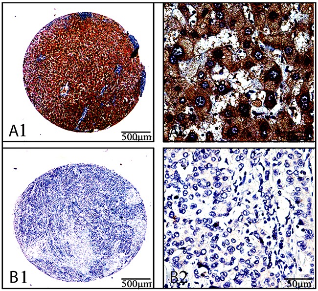
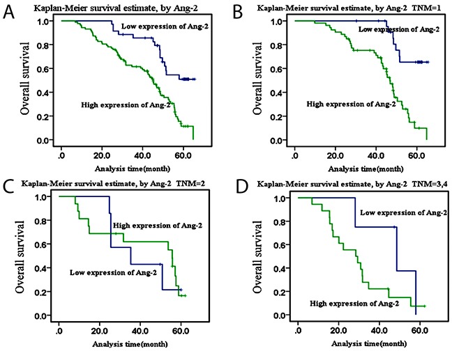
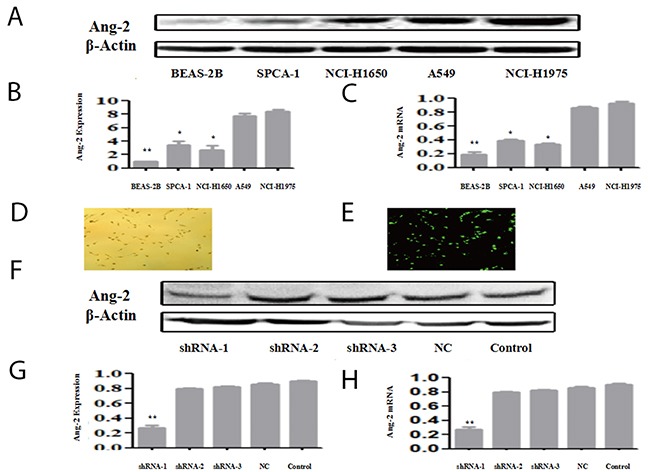
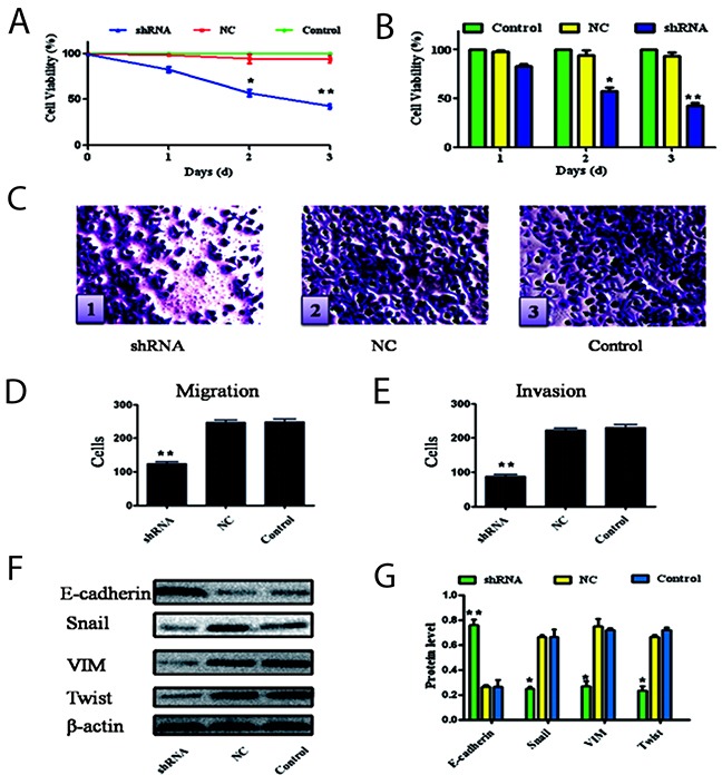
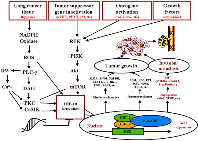
Lung cancer is the most common malignant tumor with increasing angiopoietin-2 (Ang-2) and a high rate of metastasis. However, the mechanism of Ang-2 enhancing tumor proliferation and facilitating metastasis remains to be clarified. In this study, Ang-2 expression and its gene transcription on effects of biological behaviors and epithelial-mesenchymal transition (EMT) were investigated in lung cancers. Total incidence of Ang-2 expression in the cancerous tissues was up to 91.8 % (112 of 122) with significantly higher (χ2=103.753, P2=7.883, P=0.005), differentiation degree (χ2=4.554, P=0.033), tumor node metastasis (TNM) staging (χ2=5.039, P=0.025), and 5-year survival rate (χ2 =11.220, P2=18.881, P2=0.81, P=0.776) or III & IV (χ2=1.845, P=0.174). Over-expression of Ang-2 or Ang-2 mRNA in lung A549 and NCI-H1975 cells were identified among different cell lines. When silencing Ang-2 in A549 cells with specific shRNA-1 transfection, the cell proliferation was significantly inhibited in a time-dependent manner, with up-regulating E-cadherin, down-regulating Vimentin, Twist, and Snail expression, and decreasing invasion and metastasis of cancer cell abilities, suggesting that Ang-2 promote tumor metastasis through increasing EMT, and it could be a potential target for lung cancer therapy.

Keywords: Ang-2; EMT; RNA interference; lung cancer; prognosis.


Conflict of interest statement

CONFLICTS OF INTEREST No potential conflicts of interest were disclosed.

Figures

Figure 1
Figure 1. Ang-2 expression and cellular distribution by immunohistochemistryAng-2 expression in the cancerous-and their paracancerous-tissues of 122 patients with lung cancers were analyzed by immunohistochemistry with anti-human Ang-2 antibody (ab155106, Abcam, USA)
(A1 and A2), the stronger straining of Ang-2 expression in lung adenocarcinoma tissues, with deeper brown staining particles mainly localized in the cytoplasm and cell membranes; (B1 and B2), the light straining of Ang-2 expression in their surrounding tissues; a1&b1, the original magnification × 40; a2&b2, the original magnification × 400.
Figure 2
Figure 2. Kaplan-Meier survival curves of Ang-2 overexpressionThe survival curves of high Ang-2 expression in patients with lung cancer were made by the Kaplan-Meier method
(A), the overall survival curve of high Ang-2 expression in patients with lung cancer; (B), the overall survival curve of high Ang-2 expression in patients with lung cancer at TNM stage I; (C), the overall survival curve of high Ang-2 expression in patients with lung cancer at stage II; and (D), the overall survival curve of high Ang-2 expression in patinets with lung cancer at stage III-IV, respectively.
Figure 3
Figure 3. Ang-2 expression, gene transcription and shRNA suppression Ang-2 expression and gene transcription in lung Beas-2B cell, lung cancer (SPC-A-1, NCI-1650, A549, and NCI-1975) cell lines were analyzed at protein- by Western blotting or at mRNA-level by qRT-PCR
(A), the Ang-2 expressions in different cells at protein level with β-actin as control; (B), the ratios from Ang-2 to β-actin in different cells at protein level; (C), the ratios from Ang-2 to GAPDH expression in different cells at mRNA level; (D), the phase-contrast image (X100 magnification) with the Ang-2-shRNA1 successfully transfected into A549 cells at 24 h; (E), the fluorescence image (X100 magnification) with the Ang-2-shRNA1 successfully transfected into A549 cells; (F), the different alterations of Ang-2 expression with β-actin as loading control at 48 h; (G), the ratios from Ang-2 to β-actin protein in different cell lines; and (H), the similar alterations of Ang-2 normalized to GAPDH in different cell lines at mRNA level. Ang-2, angiopoietin-2; Ang-2 Exp., angiopoietin-2 protein expression; shRNA, the Ang-2-shRNA transfection group; NC, the negative control group; and Control, the blank control group. *P<0.05. **P<0.001.
Figure 4
Figure 4. Silencing Ang-2 on effect of biological behaviors and EMT of cells
The possibilities of cell proliferation, migration, invasion and EMT of the A549 cells were altered after the most effective Ang-2-shRNA-1 transfection. (A & B), the cell proliferation ability was examined using CCK-8 at 450 nm. (C), the cell patterns (×200 magnification) with the crystal violet staining solution: c1, the cells in the shRNA group; c2, the cells in the NC group, and c3, the cells in the control group; (D), the cell migration abilities were presented as total number of cells that migrated to the bottom chamber without or with the transwell-precoated matrigel, as calculated at 6 random fields; (E), the cellinvasion abilities were presented as total number of cells that migrated to the bottom chamber without or with the transwell-precoated matrigel, as calculated at 6 random fields; (F), the EMT-related epithelial indicators and mesenchymal markers after silencing Ang-2 transcritpion of A549 cells were analyzed by the Western blotting with β-actin as loading control; and (G), the relative ratio from each protein to β-actin (n=3) compared with the control group. Ang-2, angiopoietin-2; shRNA, the Ang-2-shRNA transfection group; NC, the negative control group; and Control, the blank control group; *P<0.05. **P<0.001.
Figure 5
Figure 5. Possibility mechanism of tumor derived Ang-2 up-regulating EMT and facilitating lung cancer metastasis
Ang-2 has been implicated in mediating inflammatory processes, and upregulated in multiple inflammation-related tumors or signaling pathway. With lung cancer volume increasing, intratumoral hypoxia in the absence of vascularization resulted in tumor cells to express Ang-2 inducing angiogenesis for tumor growth and increasing vascular permeability. Overexpression of Ang-2 facilitating proliferation, invasion, metastasis, and EMT of lung cancer cells that could be a new mechanism insight into tumor metastasis by inhibiting E-cadherin and upregulating VIM, Twist and Snail signaling. Ang, angiopoietin; Ang-2, angiopoietin-2; CaMK, calcium/calmodulin-dependent protein kinase; EMT, epithelial-mesenchymal transition; ENO1, enolase 1; EPO, erythropoietin; ET1, endothelin-1; GAPDH, glyceraldehyde-3-phosphate dehydrogenase; Glut1, glucose transporter 1; HIF-1α, hypoxia-inducible factor-1α; HK1, hexokinase 1; HRE, hypoxia-response element; IGF2, insulin-like growth factor 2; IP3, inositol 1,4,5-trisphate; mTOR, mechanistic target of rapamycin kinase; NOS2, nitric oxide synthase 2; Oncogenes, ras, c-myc etc; PLC-γ, phospholipase C gamma; PI3K, phosphatidylinositol 3-kinase; PKC, protein kinase C; RTK, receptor tyrosine kinase; TGFA, transforming growth factor alpha; tumor suppressor genes, pVHL, P53, PTEN, etc. VEGF, vascular endothelial growth factors; VHL, von Hippel-Lidau; VIM, Vimentin.

Similar articles

Cited by

References

    1. Chen W, Zheng R, Zhang S, Zeng H, Zuo T, Xia C, Yang Z, He J. Cancer incidence and mortality in China in 2013: an analysis based on urbanization level. Chin J Cancer Res. 2017;29:1–10. - PMC - PubMed
    1. Malvezzi M, Carioli G, Bertuccio P, Boffetta P, Levi F, La Vecchia C, Negri E. European cancer mortality predictions for the year 2017, with focus on lung cancer. Ann Oncol. 2017;28:1117–1123. - PubMed
    1. Cassidy RJ, Zhang X, Patel PR, Shelton JW, Escott CE, Sica GL, Rossi MR, Hill CE, Steuer CE, Pillai RN, Ramalingam SS, Owonikoko TK, Behera M, et al. Next-generation sequencing and clinical outcomes of patients with lung adenocarcinoma treated with stereotactic body radiotherapy. Cancer. 2017;123:3681–3690. - PubMed
    1. Brock M, Mei Y. Protein functional effector sncRNAs (pfeRNAs) in lung cancer. Cancer Lett. 2017;403:138–143. - PubMed
    1. Lu H, Jiang Z. Advances in antiangiogenic treatment of small-cell lung cancer. Onco Targets Ther. 2017;10:353–359. - PMC - PubMed