. 2018 Feb 2;9(16):12879-12893.
doi: 10.18632/oncotarget.24387. eCollection 2018 Feb 27.

Hypoxic marker CA IX and adhesion mediator β-catenin are downregulated by lymphocytic choriomeningitis virus persistent infection

Affiliations

Hypoxic marker CA IX and adhesion mediator β-catenin are downregulated by lymphocytic choriomeningitis virus persistent infection

Andrea Fabianova et al. Oncotarget. .

Abstract

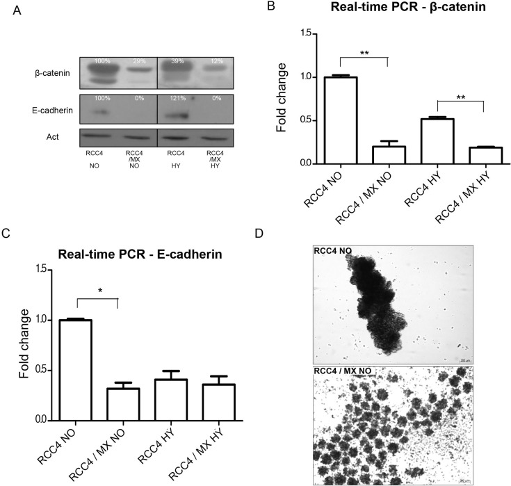
Renal cell carcinoma is one of the most frequent cancer diseases with high resistance to radio- and chemotherapy. Mutation of VHL gene is frequent in these tumors leading to simulation of hypoxic conditions. Lymphocytic choriomeningitis virus, belonging to RNA viruses, is a neglected human pathogen and teratogen. We have found that infection of renal cell carcinoma cells by lymphocytic choriomeningitis virus strain MX causes a decrease of carbonic anhydrase IX protein and RNA level. Lower expression of carbonic anhydrase IX on the cell surface provides less target for carbonic anhydrase IX-targeted immunotherapy. What more, reduced levels of adhesion mediating protein β-catenin as well as E-cadherin, as a consequence of infection, suggest a possible increase in metastatic potential of cells infected by lymphocytic choriomeningitis virus strain MX. These results might help elucidate differences in patients susceptibility to immunotherapy directed against carbonic anhydrase IX or in developing new therapeutical strategies. Our data indicate that presence of infection can significantly affect patient response to cancer therapy.

Keywords: carbonic anhydrase IX; immunotherapy; internalization; lymphocytic choriomeningitis virus; renal cell carcinoma.


Conflict of interest statement

CONFLICTS OF INTEREST The authors report no conflicts of interest.

Figures

Figure 1
Figure 1. LCMV MX strain infection of RCC4 and RCC4 VHL cell lines
(A) PCR of all viral genes, nucleoprotein (NP), Z protein (ZP), glycoprotein (GP), and L protein (LP), present in uninfected or infected RCC4 and RCC4 VHL cells (RCC4, RCC4 VHL, RCC4/MX, RCC4 VHL/MX) cultivated in normoxic (NO) or hypoxic (HY) conditions. (B) Western blot analysis for presence of viral NP in uninfected and infected RCC4 and RCC4 VHL cells cultivated in normoxic (NO) or hypoxic (HY) conditions. β-actin (Act) was used as a loading control. The numbers indicate percentage of densitometric evaluation. (C) Immunofluorescence of NP in infected or uninfected cells. Bar = 20 μm. (D) Transmitted light microscopy of uninfected RCC4 and infected RCC4/MX cells shows the changes of morphology caused by infection. Images were acquired by Microscope Zeiss Axiovert 40 CFL, magnification 200×. Bar = 50 μm. (E) Proliferation assay of RCC4 and RCC4/MX. Cells (103 cells/well) were plated into 24 well plate and counted every 12 hours. Y-axis shows the number of cells determined by cell counter at the indicated time points (X-axis).
Figure 2
Figure 2. Changes in the expression of HIF-1 targets after infection with LCMV MX or transfection with pcDNA3.1-NP
Western blot analysis was used to evaluate the levels of (A) CA IX, (C) HIF-1 α and HIF-2α and (E) HIF-1 targets, lactate dehydrogenase-A (LDH-A) and enolase (ENO) in uninfected RCC4 and infected RCC4/MX cells cultivated under normoxic (NO) and hypoxic (HY) conditions. β-actin (Act) was used as a loading control. The numbers indicate percentage of densitometric evaluation. To quantify the mRNA levels of (B) CA IX or (D) HIF-1 α in infected or control cells in normoxic (NO) and hypoxic (HY) conditions, a q-PCR with gene specific primers was done. The samples were normalized to β-actin and ΔΔCt method was used to evaluate the fold change in infected and uninfected cells. Single, double, and triple asterisk indicates p value lower than 0.05, 0.01, and 0.001, respectively, determined by the Student's t-test. (F) Western blot analysis of CA IX after transfection. Uninfected RCC4 cells were transfected by pcDNA3.1 empty vector (pcDNA3.1-EV) and pcDNA3.1-NP plasmids and subjected to western blot analysis. β-actin (Act) was used as a loading control. The numbers indicate percentage of densitometric evaluation.
Figure 3
Figure 3. Analysis of adhesion molecules expression
(A) Western blot analysis of β-catenin and E-cadherin in RCC4 and RCC4/MX cells under normoxic (NO) and hypoxic (HY) conditions. β-actin (Act) was used as a loading control. The numbers indicate percentage of densitometric evaluation. Q-PCR of (B) β-catenin and (C) E-cadherin in infected or uninfected cells cultivated under normoxic (NO) and hypoxic (HY) conditions. The samples were normalized to β-actin and ΔΔCt method was done to evaluate the fold change of infected and uninfected cells. Single and double asterisk indicates p value lower than 0.05 and 0.01, respectively, determined by the Student's t-test. (D) Aggregation assay of uninfected RCC4 and infected RCC4/MX cells. Cells (1.5 × 10⁵ cells/well) were seeded into a plate with non-adhesive surface and incubated on a rotation shaker at 120 rpm for 24 h. The cell aggregates were observed by Zeiss Axiovert 40 CFL microscope, magnification 200×. Bar = 200 μm.
Figure 4
Figure 4. CA IX localisation and internalisation of VII/20 antibody in uninfected and infected cells
(A) Determination of total level of CA IX present in uninfected and infected cells by ELISA. The total CA IX level was normalized to total amount of proteins. Y-axis shows the absorbance at OD492. Double and triple asterisk indicates p value less than 0.01 and 0.001, respectively, determined by the Student's t-test. (B) Flow cytometry of internalizing VII/20 antibody in uninfected and infected cells cultured in normoxia (NO) and hypoxia (HY). Y-axis shows fluorescence signal of VII/20 antibody. Empty bar shows total signal of the VII/20 antibody, bricked bar shows signal of only membrane CA IX bound VII/20 antibody, and dotted bar shows the signal of internalized VII/20 antibody. Double asterisk indicates p-value less than 0.01 determined by the Student's t-test.

References

    1. Southern PJ, Singh MK, Riviere Y, Jacoby DR, Buchmeier MJ, Oldstone MB. Molecular characterization of the genomic S RNA segment from lymphocytic choriomeningitis virus. Virology. 1987;157:145–55. - PubMed
    1. Wright KE, Spiro RC, Burns JW, Buchmeier MJ. Post-translational processing of the glycoproteins of lymphocytic choriomeningitis virus. Virology. 1990;177:175–83. - PMC - PubMed
    1. Salvato MS, Shimomaye EM. The completed sequence of lymphocytic choriomeningitis virus reveals a unique RNA structure and a gene for a zinc finger protein. Virology. 1989;173:1–10. - PubMed
    1. Buchmeier MJ, Welsh RM, Dutko FJ, Oldstone MB. The virology and immunobiology of lymphocytic choriomeningitis virus infection. Adv Immunol. 1980;30:275–331. - PubMed
    1. Labudova M, Tomaskova J, Skultety L, Pastorek J, Pastorekova S. The nucleoprotein of lymphocytic choriomeningitis virus facilitates spread of persistent infection through stabilization of the keratin network. J Virol. 2009;83:7842–9. https://doi.org/JVI.00309-09 - PMC - PubMed

LinkOut - more resources