. 2018 Mar 12:4:41.
doi: 10.1038/s41420-018-0041-x. eCollection 2018.

Micro RNAs are involved in activation of epicardium during zebrafish heart regeneration

Affiliations

Micro RNAs are involved in activation of epicardium during zebrafish heart regeneration

Marcello Ceci et al. Cell Death Discov. .

Abstract

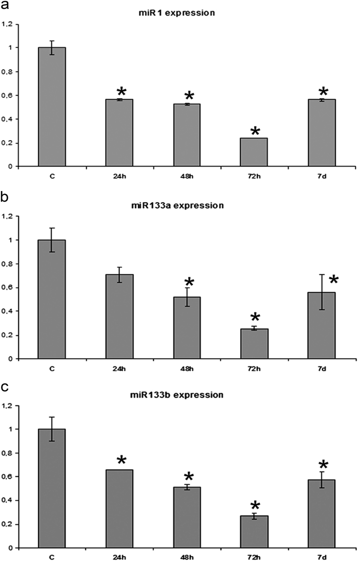
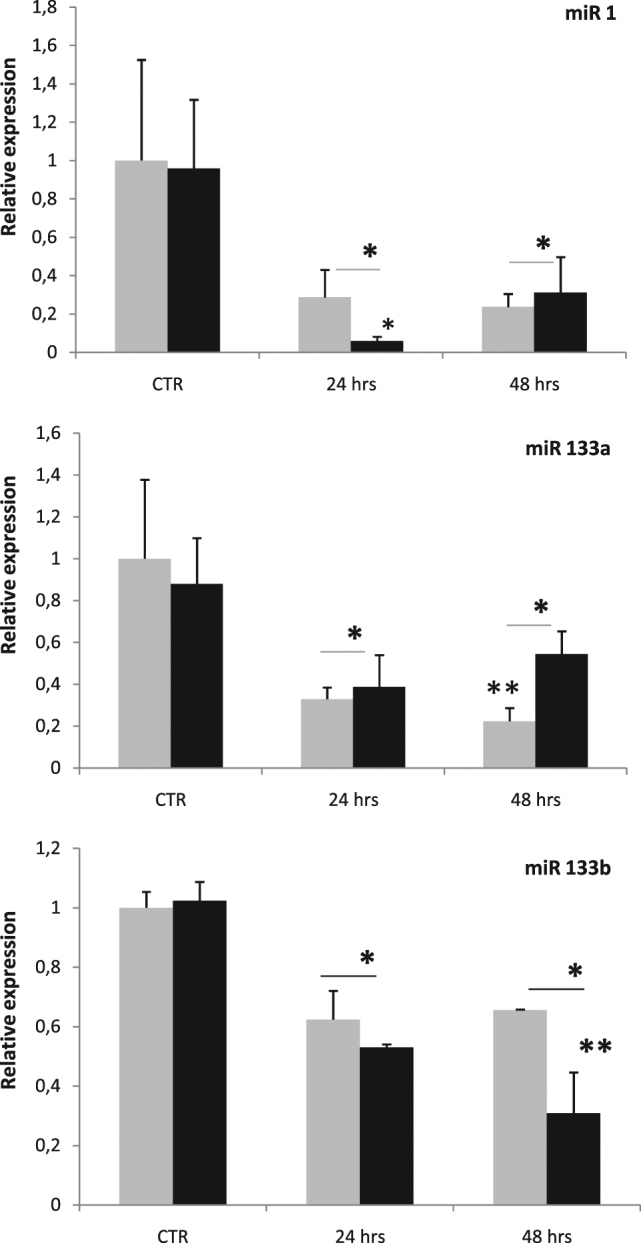
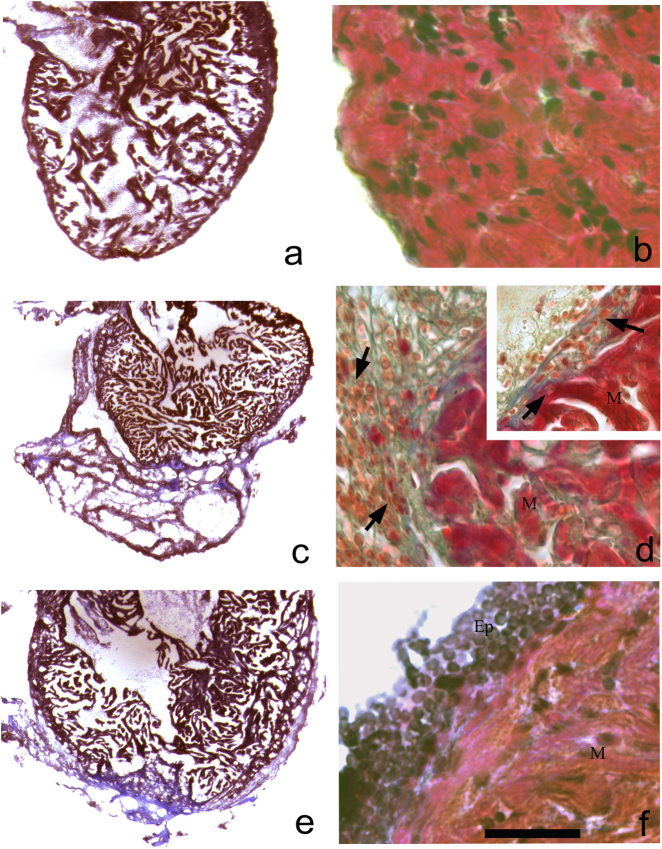
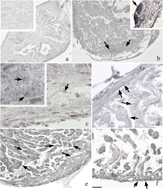
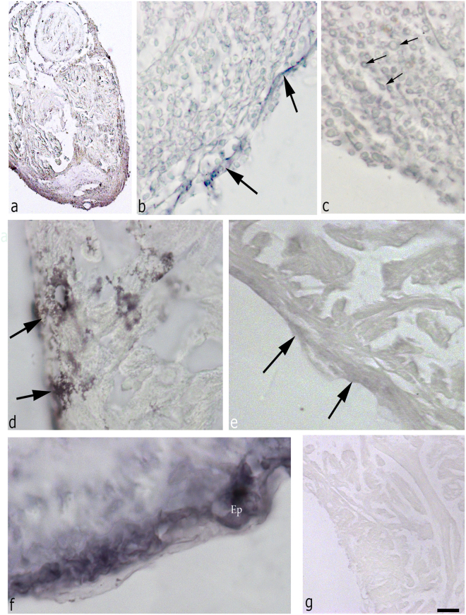
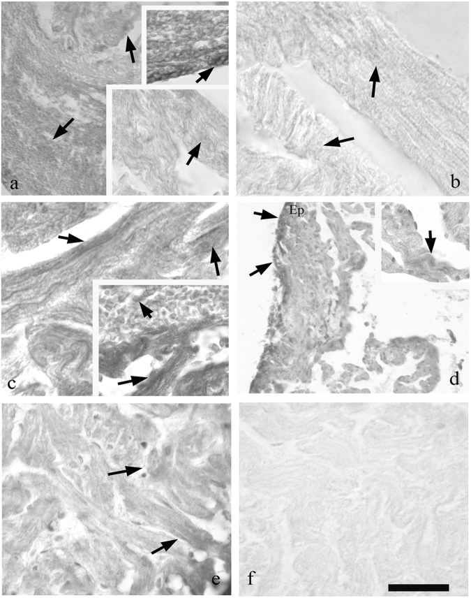
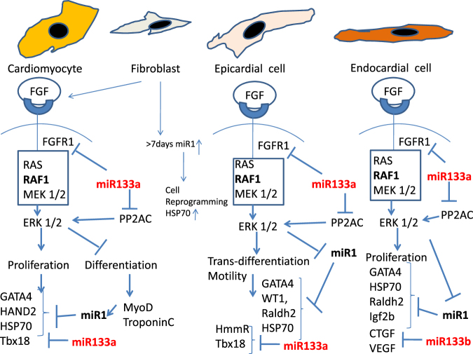
Zebrafish could be an interesting translational model to understand and improve the post-infarction trial and possible regeneration in humans. The adult zebrafish is able to regenerate efficiently after resecting nearly 20% of the ventricular apex. This process requires the concert activation of the epicardium and endocardium, as well as trans-differentiation of pre-existing cardiomyocytes that together replace the lost tissue. The molecular mechanisms involved in this activation process are not completely clarified. In this work, in order to investigate if the downregulation of these miRNAs (miRs) are linked with the activation of epicardium, the expressions of miR-133a, b and miR-1 during regeneration were analysed. qPCR analyses in whole-heart, or from distinct dissected epicardial cells comparing to regenerative clot (containing cardiomyocytes, fibroblasts and endocardial cells) by a laser-micro-dissector, have indicated that already at 24 h there is a downregulation of miRs: (1) miR-133a and miR-1 in the epicardium and (2) miR-133b and miR-1 in the regenerative clot. All the miRs remain downregulated until 7 days post-surgery. With the aim to visualize the activations of heart component in combination with miRs, we developed immunohistochemistry using antibodies directed against common markers in mammals as well as zebrafish: Wilms tumour 1 (WT1), a marker of epicardium; heat-shock protein 70 (HSP70), a chaperon activated during regeneration; and the Cardiac Troponin T (cTnT), a marker of differentiated cardiomyocytes. All these markers are directly or indirectly linked to the investigated miRs. WT1 and HSP70 strongly marked the regeneration site just at 2-3 days postventricular resection. In coherence, cTnT intensively marked the regenerative portion from 7 days onwards. miRs-1 and -133 (a,b) have been strongly involved in the activation of epicardium and regenerative clot during the regeneration process in zebrafish. This study can be a useful translational model to understand the early epicardial activation in which miRs-133a and miR-1 seem to play a central role as observed in the human heart.


Conflict of interest statement

The authors declare that they have no conflict of interest.

Figures

Fig. 1
Fig. 1
Analysis of qRT-PCR regarding the relative expression of miR in the heart during the regeneration process at 1, 2, 3 and 7 dpa. a Expression of miR-1; b expression of miR-133a; c expression of miR-133b. (*) Statistically significant difference in the expression level in comparison to control (P < 0.001) according to the Student's t-test to a queue. Normalized values with U6 snRNA
Fig. 2
Fig. 2
Quantitative PCR regarding the relative expression of miRs in the epicardial cells (EPCs, light grey) and in regenerative clot (RC, black) of adult zebrafish at 1 (24 h) and 2 (48 h) dpa. (*) Statistically significant difference in the expression level in comparison to control (P < 0.001) according to the Student's t-test to a queue. Normalized values with U6 snRNA. (**) Values statistically significant (P < 0.001) EPCs vs RC
Fig. 3
Fig. 3
Histological staining using Masson’s trichrome on heart controls (a, b) and during regeneration (c–f). At 2 dpa (c) wide regenerative clot is clearly evidenced; in d a fibrous clot in blue is observed, while some undifferentiated cells that infiltrated the clot-surrounded epicardium are reactive (d, inset). We also notice some macrophages (d, inset). A 3 days (e) the clot is in resorption; in a magnification (f) epicardial stratification is observed. M myocytes, Ep epicardium (a bar = 1.5 mm; b bar = 45 μm; c, e bar = 1.5 mm; d, f bar = 45 μm)
Fig. 4
Fig. 4
Immunohistochemistry with HSP70 on zebrafish heart. a HSP70 staining in the control. The reaction is marked in the samples of 2 and 3 dpa (b and d, respectively); in particular in the regions of the clot (c, and inset) and epicardium (Ep) (b, inset). In the region of cutting are visible at 2 dpa many rounded positive cells. From 14 days (f) onwards, the clot has almost disappeared and replaced by new myocytes (M); is visible to the evidenced several erythrocytes in the heart lumen (e). At days 30, (f) positivity is still reactive than in the control (a bar = 300 μm; inset = 75 μm; b bar = 300 μm and inset bar = 4 μm; c bar = 75 μm and inset bar = 45 μm; d bar = 150 μm and bar = 150 μm; f bar = 75 μm)
Fig. 5
Fig. 5
Immunohistochemistry with WT1 on regenerative heart (a bar = 300 µm; b bar = 150 µm; c bar = 75 µm; d bar = 45 µm; e bar = 50 µm; f bar = 45 µm; g bar = 150 µm). At 2 days, (a) there is a strong reactivity in the epicardium surrounding the organ. This positivity is present also in EPCs surrounding the clot (b) and in cells inside the clot (c). At 3 dpa, (d) the epicardium is reactive, but at 7 dpa (e) the signal in some epicardial portions is lower as compared with the parts next to the RC (f). In g, the control staining (bar = 75 µm). Ep epicardium
Fig. 6
Fig. 6
Immunohistochemistry with cTnT on regenerating heart (a, e lower bar = 75 μm and upper inset bar = 45 μm; b bar = 54 μm; c bar = 45 μm and inset bar = 54 μm; d, bar = 150 μm and inset bar = 75 μm; and bar = 54 μm). At 2 days (a) are observed healthy muscle fibres with point reactivity (inset bottom) and highly reactive rounded cells (upper inset) among the heatly fibres. At 3 (b) and 7 (c) dpa next to healthy fibres (M) cTnT+ cells are evidenced close the clot (c, inset). As is shown in 14 days, (d) the epicardium (Ep) is cTnT posititve; infiltrated cells in the clot are also positive. At 30 dpa, (e) muscle fibres are reformed in the region of the cut and the reactivity for cTnT is comparable to the control. In f is the sample stained with omission of cTnT that shows no reactivity
Fig. 7
Fig. 7
A proposed schematic model of miR-1 and miR-133 actions in blocking the FGF-dependent transduction pathway in the cells involved in cardiac regeneration: CMs, fibroblast, EPCs and endocardial cells. In zebrafish these four types acting in concert, the regeneration in trans-differentiation and/or proliferation of existing resident cells under the induction of FGF1-2. The scheme is a comprehensive information from literature and from the present research. miR-1 is not expressed by ERK1,2 activity because of the expression of cardiac embryonal genes and relative proteins such as GATA4. When the genes of differentiation are expressed, such as MyoD, miR-1 start to be highly transcripted and act as a repressor of GATA4 translation and other embryonic key proteins. For example, in epicardial cells it may control the WT1 expression. miR-133a instead acts directly on blocking the expression of the receptor of FGF and on the expression of the PP2AC that promotes the activity of ERK. miR-133b is instead involved in the transition from endothelial to mesenchymal cells by blocking directly the expression of connective tissue growth factor (CTGF)

References

    1. Kikuchi K. Advances in understanding the mechanism of zebrafish heart regeneration. Stem Cell Res. 2014;13:542–555. doi: 10.1016/j.scr.2014.07.003. - DOI - PubMed
    1. Chimenti I, et al. Cardiosphere-derived cells transplanted into infarcted mice relative roles of direct regeneration versus paracrine effects of human. Circ. Res. 2010;106:971–980. doi: 10.1161/CIRCRESAHA.109.210682. - DOI - PMC - PubMed
    1. Wu C, Weidinger G. Zebrafish as a model for studying cardiac regeneration. Curr. Pathobiol. Rep. 2014;2:93–100. doi: 10.1007/s40139-014-0042-2. - DOI
    1. Poss KD, Wilson LG, Keating MT. Heart regeneration in zebrafish. Science. 2002;298:2188–90. doi: 10.1126/science.1077857. - DOI - PubMed
    1. Lepilina A, et al. A dynamic epicardial injury response supports progenitor cell activity during zebrafish heart regeneration. Cell. 2006;127:607–619. doi: 10.1016/j.cell.2006.08.052. - DOI - PubMed

LinkOut - more resources