. 2018 Mar 12:4:42.
doi: 10.1038/s41420-018-0040-y. eCollection 2018.

Regulation of transforming growth factor is involved in the efficacy of combined 5-fluorouracil and interferon alpha-2b therapy of advanced hepatocellular carcinoma

Affiliations

Regulation of transforming growth factor is involved in the efficacy of combined 5-fluorouracil and interferon alpha-2b therapy of advanced hepatocellular carcinoma

Youhei Okada et al. Cell Death Discov. .

Abstract

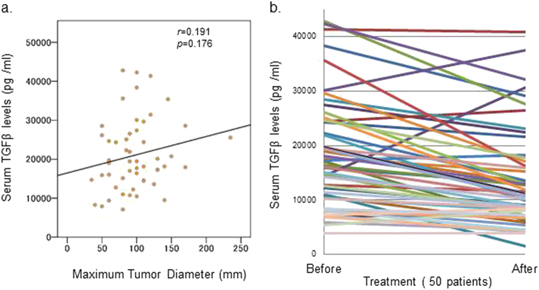
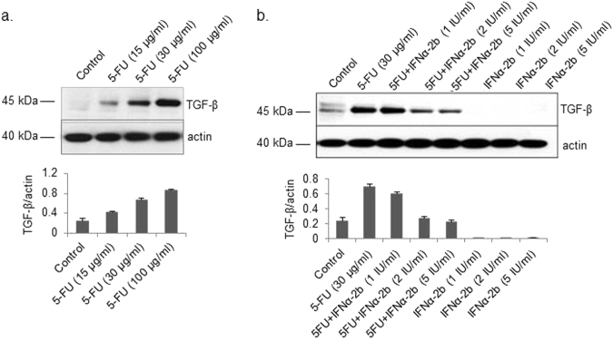
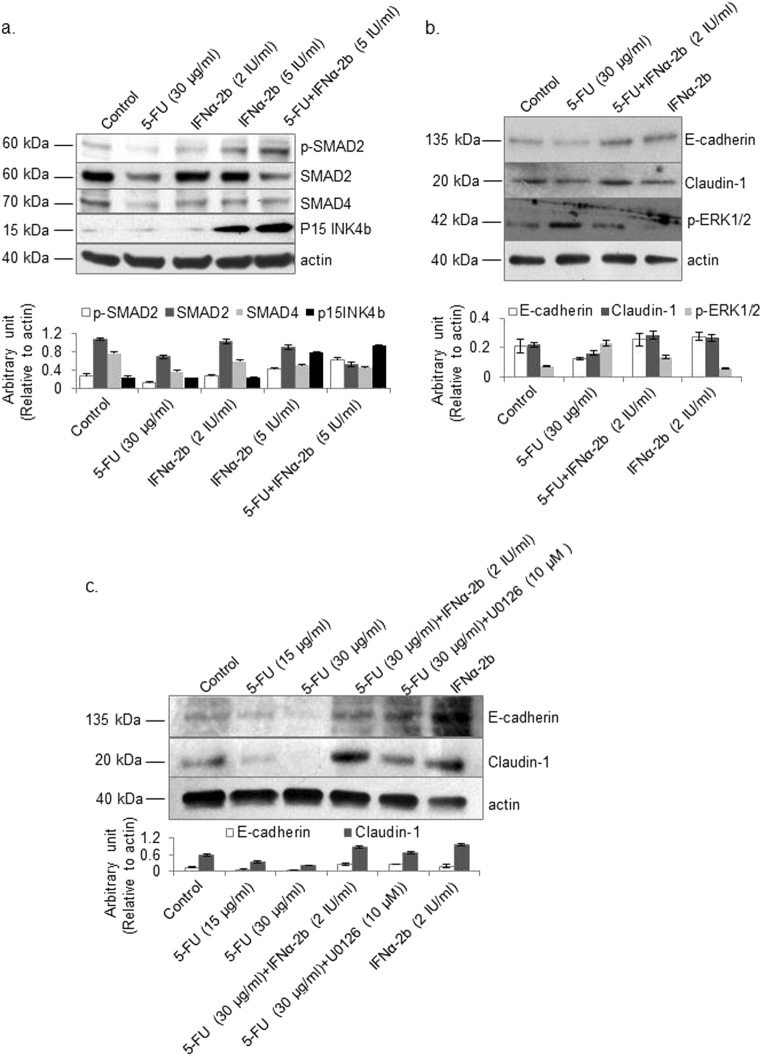
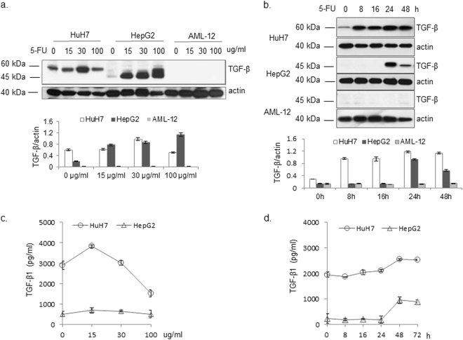
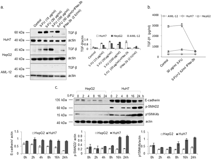
Transforming growth factor-beta (TGF-β) is critical in cancer cell invasion and metastasis. The effects of a treatment that targets TGF-β using the combination of interferon alpha (IFNα)-2b and 5-fluorouracil (5-FU) are unknown. Here, we show that the serum levels of TGF-β1 prior to the therapy correlate with increased maximum tumor diameter, which is significantly (p < 0.01) decreased after the combination therapy. 5-FU increased both the expression and secretion levels of TGF-β1 in hepatoma cells, but not in normal hepatocytes. The combination of 5-FU and IFNα-2b synergistically affected cell death. However, a TGF-β1 specific inhibitor did not affect the anti-tumor activity of 5-FU. 5-FU inhibited the phosphorylation of SMAD2 and reduced the total protein levels of SMAD2, SMAD4, and pINK4b. Conversely, 5-FU stimulated the phosphorylation of extracellular signal-regulated kinase (ERK)1/2. Accordingly, the protein levels of E-cadherin and claudin-1 were reduced in 5-FU-treated cells. The combination of 5-FU and IFNα-2b, and the inhibition of ERK1/2 by a specific inhibitor neutralized the effects of 5-FU on TGF-β-related signaling molecules and restored their protein levels to those observed in the control. Interestingly, the phosphorylated protein levels of SMAD2 and the total protein levels of E-cadherin and p15INK4b were increased in 5-FU-stimulated HuH-7 cells, but not in Hep G2 cells. Our data suggest that the higher efficacy of the 5-FU and IFNα-2b combination therapy was associated with the regulation of TGF-β expression, secretion, and the signals mediated by it.


Conflict of interest statement

The authors declare that they have no conflict of interest.

Figures

Fig. 1
Fig. 1. Detection of TGF-β1 serum levels in patients with advanced HCC.
a TGF-β1 levels before the treatment were used to analyze the correlation with the maximum tumor diameter. A p-value and its correlation were calculated in Microsoft Excel 2010. b TGF-β1 serum levels in patients before and after treatment were determined according to the manufacturer’s instructions. Fifty patients were analyzed. Statistical comparisons were performed using a paired t-test. A p-value < 0.05 was considered to be significant
Fig. 2
Fig. 2. Effects of 5-FU and IFNα-2b alone and in combination on TGF-β levels in Hep G2 cells.
Cells were treated with 0, 15, 30, and 100 µg/mL 5-FU for 24 h (a) or with 30 µg/mL 5-FU, IFNα-2b (1, 2, 5 IU/mL), or 5-FU and IFNα-2b for 24 h (b). TGFβ levels in the cell lysates were determined by western blotting. Results are expressed as the means ± SD (n = 3)
Fig. 3
Fig. 3. Effects of 5-FU treatment alone, or in combination with IFNα-2b or TGF-β receptor inhibitor, on the viability and apoptosis of Hep G2 cells.
a Cells were treated with 15 µg/mL 5-FU, 2 IU/mL IFNα-2b, or a combination of both for 48 h. In additional experiments, cells were pretreated with 10 µM SB-431542 (a TGF-β inhibitor) before treatment with 5-FU. The number of viable cells was evaluated by checking the optical density at 450 nm. b Cells were given the same treatment as described in a; thereafter, the apoptotic cells were stained, photographed (scale = 400 μM), and counted (c) as described in the materials and methods section. The statistical analysis was carried out using one-way ANOVA-POST HOC (Tukey’s HSD) analysis. A p-value < 0.05 was considered to be significant. Results are expressed as the means ± SD (n = 3)
Fig. 4
Fig. 4. Effects of 5-FU and IFNα-2b alone and in combination on TGF-β-mediated signaling.
Hep G2 cells were treated with 5-FU and/or IFNα-2b at the indicated concentrations for 24 h. a The protein levels of p-SMAD2, SMAD2, SMAD4, and p15INK4b were detected by western blotting. b In another experiment, levels of EMT molecules including E-cadherin, claudin-1, and p-ERK1/2 were detected by western blotting as described in the materials and methods section. c Besides the treatment of 5-FU and IFNα-2b alone and in combination, the ERK1/2 inhibitor U0126 was used to clarify the role of ERK1/2 in the regulation of E-cadherin and claudin-1 by 5-FU. Actin was detected as an internal control. The quantitative data are presented as the means ± SD (n = 3)
Fig. 5
Fig. 5. Cell-dependent regulation of TGF-β expression and secretion by 5-FU.
HuH-7, Hep G2, and AML-12 cells were treated with different concentrations of 5-FU as indicated (a), and 30 µg/mL 5-FU for the indicated time periods (b). TGF-β was detected by western blotting. TGF-β secretion was measured in HuH-7 and Hep G2 cells as described in Materials and Methods (c, d). Results are expressed as the means ± SD (n = 3)
Fig. 6
Fig. 6. Responses of different cells to 5-FU, IFNα-2b, and combined treatment with respect to TGF-β levels and TGF-β-mediated signaling.
Hep G2, HuH-7, and AML-12 cells were individually treated with 5-FU, IFNα-2b, and a combination of 5-FU and IFNα-2b for 24 h. TGF-β levels in cell lysates (a) and culture supernatants (b) were detected. (c) Both Hep G2 and HuH-7 cells were treated with 5-FU for different periods as indicated. The levels of E-cadherin, p-SMAD2, and p15INK4b were detected by western blot. Actin was detected as an internal control. Results are expressed as the means ± SD (n = 3)

Similar articles

Cited by

References

    1. Schwartz M, Roayaie S, Konstadoulakis M. Strategies for the management of hepatocellular carcinoma. Nat. Clin. Pract. Oncol. 2007;4:424–432. doi: 10.1038/ncponc0844. - DOI - PubMed
    1. Sakon M, et al. Combined intraarterial 5-fluorouracil and subcutaneous interferon-alpha therapy for advanced hepatocellular carcinoma with tumor thrombi in the major portal branches. Cancer. 2002;94:435–442. doi: 10.1002/cncr.10246. - DOI - PubMed
    1. Obi S, et al. Combination therapy of intraarterial 5-fluorouracil and systemic interferon-alpha for advanced hepatocellular carcinoma with portal venous invasion. Cancer. 2006;106:1990–1997. doi: 10.1002/cncr.21832. - DOI - PubMed
    1. Kasai K, et al. Therapeutic efficacy of combination therapy with intra-arterial 5-fluorouracil and systemic pegylated interferon alpha-2b for advanced hepatocellular carcinoma with portal venous invasion. Cancer. 2012;118:3302–3310. doi: 10.1002/cncr.26648. - DOI - PubMed
    1. Kasai K, et al. Combination therapy of intra-arterial 5-fluorouracil and systemic pegylated interferon alpha-2b for advanced hepatocellular carcinoma. Int. J. Clin. Oncol. 2011;16:221–229. doi: 10.1007/s10147-010-0151-9. - DOI - PubMed