. 2018 Mar 12:3:10.
doi: 10.1038/s41541-018-0048-6. eCollection 2018.

Vaccination route can significantly alter the innate lymphoid cell subsets: a feedback between IL-13 and IFN-γ

Affiliations

Vaccination route can significantly alter the innate lymphoid cell subsets: a feedback between IL-13 and IFN-γ

Zheyi Li et al. NPJ Vaccines. .

Abstract

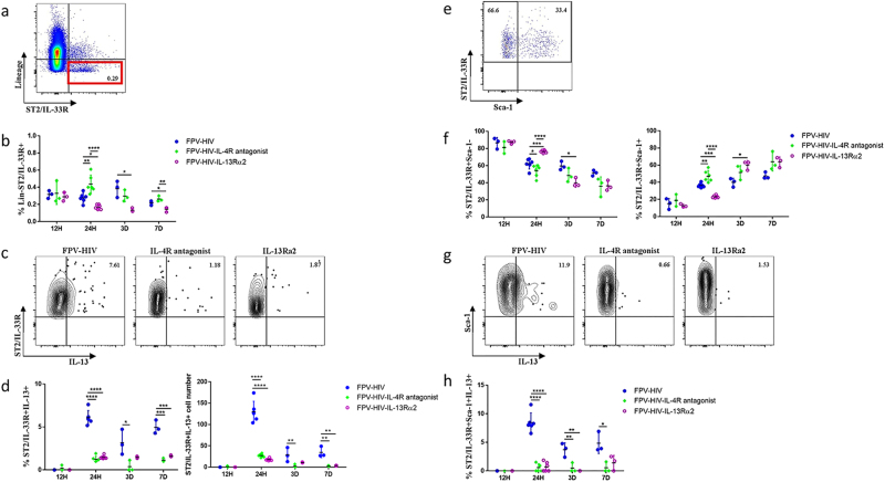
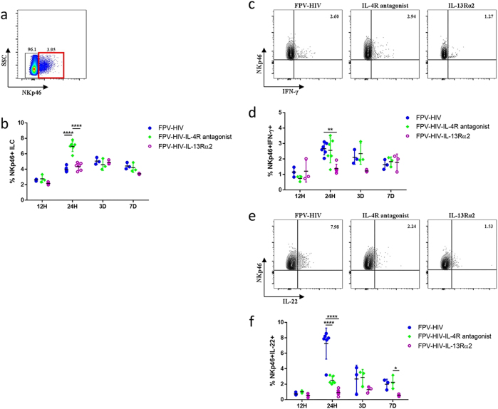
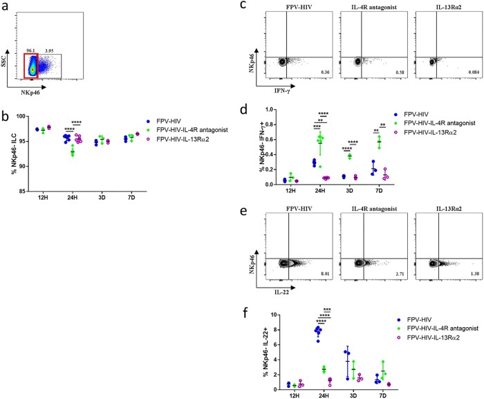
This study demonstrates that the fate of a vaccine is influenced by the cytokines produced by the innate lymphoid cells (ILC) recruited to the vaccination site, and it is vaccine route and adjuvant dependent. Intranasal virus vaccination induced ST2/IL-33R+ ILC2 in lung, while intramuscular vaccination induced exclusively IL-25R+ ILC2 in muscle. Interestingly, a larger proportion of IL-13+ ILC2s were detected in muscle following i.m. viral vector vaccination compared to lung post i.n. delivery. These observations revealed that ILC2 were the main source of IL-13 at the vaccination site (24 h post vaccination) responsible for inducing T cells of varying avidities. Moreover, recombinant fowlpox viral vector-based vaccines expressing adjuvants that transiently block IL-13 signalling at the vaccination site using different mechanisms (IL-4R antagonist or IL-13Rα2 adjuvants), revealed that the level of IL-13 present in the milieu also significantly influenced IFN-γ, IL-22 or IL-17A expression by ILC1/ILC3. Specifically, an early IL-13 and IFN-γ co-dependency at the ILC level may also be associated with shaping the downstream antibody responses, supporting the notion that differentially regulating IL-13 signalling via STAT6 or IL-13Rα2 pathways can modify ILC function and the resulting adaptive T- and B-cell immune outcomes reported previously. Moreover, unlike chronic inflammatory or experimentally induced conditions, viral vector vaccination induced uniquely different ILC profiles (i.e., expression of CD127 only on ILC2 not ILC1/ILC3; expression of IFN-γ in both NKP46+ and NKp46- ILCs). Collectively, our data highlight that tailoring a vaccine vector/adjuvant to modulate the ILC cytokine profile according to the target pathogen, may help design more efficacious vaccines in the future.


Conflict of interest statement

The authors declare no competing financial interests.

Figures

Fig. 1
Fig. 1
Evaluation of lung ILC2 and IL-13 expression following intranasal vaccination. In this study, ILC2 cells were identified as CD45+ FSClow SSClow lineageST2/IL-33R+ cells using flow cytometry (a). BALB/c mice were immunised intranasally with FPV-HIV, FPV-HIV-IL-4 antagonist and FPV-HIV-IL-13Rα2 vaccines, and the percentage of lung lineage ST2/IL-33R+ ILC2 (b) and their IL-13 expression (c, d) were assessed at 12 h, 24 h, 3 days, and 7 days post vaccination. The FACS plots (c) are representative of the 24 h data points for each vaccination. d The left graph represents percentage of ST2/IL-33R+ IL-13+ cells and right graph the total cell number. The lineage- ST2/IL-33R+ cells were further analysed for Sca-1 and IL-13 expression (eh). Data indicate that IL-13 was only detected in lineage- ST2/IL-33R+ Sca-1+ ILC2 while no IL-13 was detected in lineage- ST2/IL-33R+ Sca-1- ILC2 (gh). The graphs represent the mean and standard deviation (s.d.). The p-values were calculated using GraphPad Prism software (version 6.05 for Windows). *p < 0.05, **p < 0.01, ***p < 0.001, ****p < 0.0001. For each time point, experiments were repeated minimum three times. Note that during early time points, no significant inflammatory infiltrates were detected and the absolute cell numbers obtained were very similar
Fig. 2
Fig. 2
Expression of IFN-γ and IL-22 by NKp46+ ILC following i.n. rFPV immunisation. BALB/c mice were immunised intranasally with FPV-HIV, FPV-HIV-IL-4 antagonist, and FPV-HIV-IL-13Rα2 vaccines and the percentage of lung NKp46+ ILC cells were evaluated at 12 h, 24 h, 3 days, and 7 days post vaccination. Cells were gated as CD45+, FSClow, SSClow, lineage, ST2/IL-33R NKp46+ (a, b), and IFN-γ (c, d) and IL-22 (e, f) expression were evaluated using intracellular cytokine staining. The graphs represent the mean and standard deviation (s.d.). The p-values were calculated using GraphPad Prism software (version 6.05 for Windows). *p < 0.05, **p < 0.01, ****p < 0.0001 (one-way ANOVA). For each time point, experiments were repeated minimum three times
Fig. 3
Fig. 3
Expression of IFN-γ and IL-22 by NKp46 ILC cells post rFPV immunisation. BALB/c mice were immunised intranasally with FPV-HIV, FPV-HIV-IL-4 antagonist, and FPV-HIV-IL-13Rα2 vaccines and the percentage of lung NKp46 ILC cells were evaluated at 12 h, 24 h, 3 days, and 7 days post vaccination. Cells were gated as CD45+, FSClow, SSClow, lineage, ST2/IL-33R NKp46 (a, b), and IFN-γ (c, d) and IL-22 (e, f) expression were evaluated using intracellular cytokine staining. The graphs represent the mean and standard deviation (s.d.). The p-values were calculated using GraphPad Prism software (version 6.05 for Windows). *p < 0.05, **p < 0.01, ****p < 0.0001 (one-way ANOVA). For each time point experiments were repeated minimum three times
Fig. 4
Fig. 4
Evaluation of ILC2s and their IL-13 expression in the quadriceps muscle 24 h post intramuscular vaccination. BALB/c mice were immunised intramuscularly with FPV-HIV, FPV-HIV-IL-4 antagonist, and FPV-HIV-IL-13Rα2 vaccines and the percentage of ILC2 in muscle cells were evaluated 24 h post vaccination (note that no ST2/IL-33R+ ILC2 were detected in muscle). In this study, ILC2 cells were identified as CD45+ FSClow SSClow lineage ST2/IL-33R IL-25R+ cells using flow cytometry (a, b). The FACS plots are representative of each vaccination (b) and the collected data are presented in graph (c). The expression of IL-13 by IL-25R+ ILC2s were also assessed (d, e). The graphs represent mean and standard deviation (s.d.). The p-values were calculated using GraphPad Prism software (version 6.05 for Windows). *p < 0.05, **p < 0.01, ****P < 0.0001 (one-way ANOVA). For each time point, experiments were repeated three times. See Supplementary Fig. S1c for gating strategy
Fig. 5
Fig. 5
Evaluation of NKp46+/- ILC cytokines expression in the quadriceps muscle 24 h post intramuscular vaccination. BALB/c mice were immunised intramuscularly with FPV-HIV, FPV-HIV-IL-4 antagonist, and FPV-HIV-IL-13Rα2 vaccines, and the percentage of NKp46+ and NKp46 ILC in muscle cells were evaluated 24 h post vaccination. The NKp46+ and NKp46 ILCs were evaluated exactly as per lung tissue (a). Data represent percentage NKp46and/or NKp46+ cells expressing IFN-γ (b), IL-22 (c, d) and IL-17A (e, f). The graphs represent the mean and standard deviation (s.d.). The p-values were calculated using GraphPad Prism software (version 6.05 for Windows). *p < 0.05, **p < 0.01, ****p < 0.0001 (one-way ANOVA). For each time point experiments were repeated three times

Similar articles

Cited by

References

    1. Artis D, Spits H. The biology of innate lymphoid cells. Nature. 2015;517:293–301. doi: 10.1038/nature14189. - DOI - PubMed
    1. Sanos SL, Diefenbach A. Innate lymphoid cells: from border protection to the initiation of inflammatory diseases. Immunol. Cell Biol. 2013;91:215–224. doi: 10.1038/icb.2013.3. - DOI - PubMed
    1. Spits H, Di Santo JP. The expanding family of innate lymphoid cells: regulators and effectors of immunity and tissue remodeling. Nat. Immunol. 2011;12:21–27. doi: 10.1038/ni.1962. - DOI - PubMed
    1. Monticelli LA, et al. Innate lymphoid cells promote lung-tissue homeostasis after infection with influenza virus. Nat. Immunol. 2011;12:1045–1054. doi: 10.1038/ni.2131. - DOI - PMC - PubMed
    1. Licona-Limon P, Kim LK, Palm NW, Flavell RA. TH2, allergy and group 2 innate lymphoid cells. Nat. Immunol. 2013;14:536–542. doi: 10.1038/ni.2617. - DOI - PubMed