. 2018 Mar 16:3:7.
doi: 10.1038/s41392-017-0004-3. eCollection 2018.

Controlled drug delivery vehicles for cancer treatment and their performance

Affiliations

Controlled drug delivery vehicles for cancer treatment and their performance

Sudipta Senapati et al. Signal Transduct Target Ther. .

Abstract

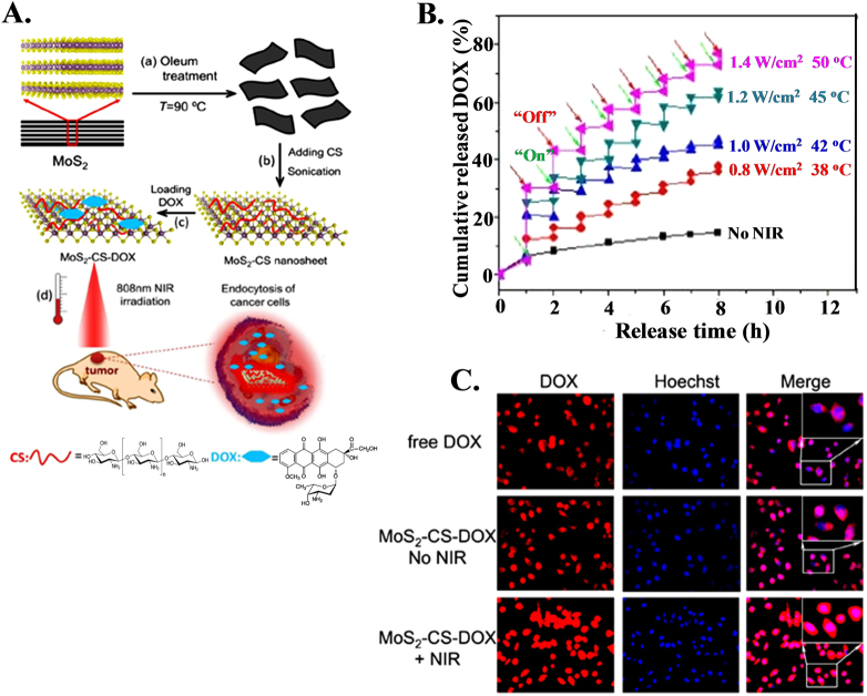
Although conventional chemotherapy has been successful to some extent, the main drawbacks of chemotherapy are its poor bioavailability, high-dose requirements, adverse side effects, low therapeutic indices, development of multiple drug resistance, and non-specific targeting. The main aim in the development of drug delivery vehicles is to successfully address these delivery-related problems and carry drugs to the desired sites of therapeutic action while reducing adverse side effects. In this review, we will discuss the different types of materials used as delivery vehicles for chemotherapeutic agents and their structural characteristics that improve the therapeutic efficacy of their drugs and will describe recent scientific advances in the area of chemotherapy, emphasizing challenges in cancer treatments.


Conflict of interest statement

The authors declare that they have no conflict of interest.

Figures

Scheme 1
Scheme 1
Different types of nanocarriers used as controlled delivery vehicles for cancer treatment
Fig. 1
Fig. 1
In vitro and in vivo controlled release of drug using layered double hydroxides and its effects. a In vitro drug release profiles for drug intercalated nitrate, carbonate and phosphate LDHs (LN-R, LC-R and LP-R, respectively); inset figure describes the release pattern of the above mentioned systems in a time frame of 0–8 h; b In vitro cytotoxicity of free drug and drug intercalated LDHs against HeLa cells at different time intervals; c In vivo antitumor effect and systematic toxicity of pure RH and drug intercalated LDHs in comparison to control; and d Histological analysis of liver, kidney and spleen of tumor bearing mice treated with control (saline), pure RH, LN-R and LP-R
Fig. 2
Fig. 2
Effect of surface modification on magnetic nanoparticle on hypothermia to reduce tumor size. a Schematic presentation showing the composition of the 4-tetracarboxyphenyl porphyrin (TCPP)-labeled, dopamine-anchored tetraethylene glycol ligands coated bimagnetic Fe/Fe3O4 nanoparticles; b Graph illustrating the temperature profiles at the MNP injection site in the body core during alternating magnetic field (AMF) exposure, which is measured with a fiber optic temperature probe; c In vivo antitumor response after intratumoral injection of MNPs followed by AMF treatments. Graph demonstrates the relative changes in average tumor volumes over time of B16–F10 tumor bearing mice that were later injected with either saline or MNP intratumorally with or without AMF treatments
Fig. 3
Fig. 3
Co-asembly of drug and photo photosensitizer for better imaging of tumor size during treatment. a Schematic representation of carrier-free nanoparticles (NPs) via co-assembly between DOX and Ce6; b In vivo fluorescence images of free Ce6 solution and Dox/Ce6 nanoparticles (NPs). The areas in the black circles represent tumor tissue; c Representative ex vivo fluorescence imaging of tumor and organs excised from Balb/c nude mice xenografted MCF-7 tumor at 24 h post-injection
Fig. 4
Fig. 4
Control delivery of drug using hydrogel as vehicle. a Illustration of the preparation and drug release of Salecan/PMAA semi-IPN hydrogels; b In vitro Dox release behaviors from the semi-IPN sample at two different pH values of 5.0 and 7.4; c Fluorescent microscopy images of A549 and HepG2 cells after 4 h of incubation with 6 μg/mL free Dox solutions and the extract liquid of Dox-loaded hydrogel; d Intravital real-time fluorescence images of ICR mice injected with FITC-labeled PMAA nanohydrogels
Fig. 5
Fig. 5
Stimuli-responsive targeted delivery of therapeutic agents. a Schematic illustration of stimuli-responsive DDS; b Schematic diagram of pH and GSH dual-responsive dynamic crosslinked supramolecular network on MSN-SS-(EDA-PGOHMA) and synthetic route with CB assembly; c Design of temperature-sensitive liposomes composed of thermosensitive poly(EOEOVE)-OD4 (i), membrane-forming EYPC (ii), membrane-stabilizing cholesterol (iii), and highly hydrophilic and nontoxic PEG-lipid (iv). Heat-triggered release of DOX from liposomes is illustrated with the structure of poly(EOEOVE)-OD4
Fig. 6
Fig. 6
Reslease and cellular uptake of drug using magnetic nanoparticles under magnetic field. a Schematic representation of a four armed PE−PCL immobilized magnetic nanoparticle (MNP); b Schematic representation of DOX-loaded MNP and DOX release under the influence of high field alternating magnetic field (HFAMF); c The release kinetics of MNP 3 (particle size of 3 nm) and MNP 5 (particle size of 5 nm) under the influence of HFAMF at 37 °C; d Cellular uptake study of DOX-loaded MAPM on HeLa cell in the presence of a static magnetic field where the nucleus was stained by DAPI (blue). The scale bar is 40 μm
Fig. 7
Fig. 7
Electric field guided control release of drug. a General scheme for the application of this system: (i) the nanoparticle-polymer solution is (ii) subcutaneously injected into a mouse, followed by (iii) application of a DC electric field to induce the release of drug cargo inside the nanoparticles; b Released amount of daunorubicin in PBS (pH 7.2) following an applied voltage (0.5 V) duration of 10 s, repeated every 5 min
Fig. 8
Fig. 8
Laser guided control drug delivery using MoS2 for cancer treatment. a Schematic illustration of high-throughput synthesis of MoS2-CS nanosheets as an NIR photothermal-triggered drug delivery system for efficient cancer therapy. (i, ii) Oleum treatment exfoliation process to produce single-layer MoS2 nanosheets that are then modified with CS, (iii) DOX loading process, and (iv) NIR photothermal-triggered drug delivery of the MoS2 nanosheets to the tumor site. b Release profile of DOX in PBS buffer (pH 5.00) in the absence and presence of an 808-nm NIR laser. c Fluorescence images of KB cells treated with free DOX, MoS2-CS-DOX, and MoS2-CS-DOX under 808-nm NIR irradiation (inset: high magnification of the rectangle area)

Similar articles

Cited by

References

    1. Boyle P, Bernard L. World Cancer Report 2008. Lyon: IARC Press; 2008.
    1. Soppimath KS, Aminabhavi TM, Kulkarni AR, Rudzinski WE. Biodegradable polymeric nanoparticles as drug delivery devices. J. Control. Release. 2001;70:1–20. doi: 10.1016/S0168-3659(00)00339-4. - DOI - PubMed
    1. Su J, Chen F, Cryns VL, Messersmith PB. Catechol polymers for pH-responsive, targeted drug delivery to cancer cells. J. Am. Chem. Soc. 2011;133:11850–11853. doi: 10.1021/ja203077x. - DOI - PMC - PubMed
    1. Kumar S, et al. Controlled drug release through regulated biodegradation of poly(lactic acid) using inorganic salts. Int. J. Biol. Macromol. 2017;104:487–497. doi: 10.1016/j.ijbiomac.2017.06.033. - DOI - PubMed
    1. Shim MS, Kwon YJ. Stimuli-responsive polymers and nanomaterials for gene delivery and imaging applications. Adv. Drug Deliv. Rev. 2012;64:1046–1059. doi: 10.1016/j.addr.2012.01.018. - DOI - PubMed

Publication types